
Uterine didelphys with three consecutive spontaneous pregnancies occurring in different side of the hemi-uterus and delivered at term by caesarean sections: a case report
Adegoriola Ojurongbe, Oluwasegun Akanni, Matthew Olusegun Fijabiyi, William Taiwo, Patrick Kudaisi
Corresponding author: Matthew Olusegun Fijabiyi, Department of Obstetrics Gynaecology, College of Clinical Sciences, Ladoke Akintola University of Technology, Ogbomoso, Oyo State, Nigeria 
Received: 12 Nov 2022 - Accepted: 14 Jan 2023 - Published: 16 Jan 2023
Domain: Obstetrics and gynecology
Keywords: Uterine didelphys, spontaneous pregnancies, term pregnancy, caesarean section, case report
©Adegoriola Ojurongbe et al. PAMJ Clinical Medicine (ISSN: 2707-2797). This is an Open Access article distributed under the terms of the Creative Commons Attribution International 4.0 License (https://creativecommons.org/licenses/by/4.0/), which permits unrestricted use, distribution, and reproduction in any medium, provided the original work is properly cited.
Cite this article: Adegoriola Ojurongbe et al. Uterine didelphys with three consecutive spontaneous pregnancies occurring in different side of the hemi-uterus and delivered at term by caesarean sections: a case report. PAMJ Clinical Medicine. 2023;11:20. [doi: 10.11604/pamj-cm.2023.11.20.38186]
Available online at: https://www.clinical-medicine.panafrican-med-journal.com//content/article/11/20/full
Case report 

Uterine didelphys with three consecutive spontaneous pregnancies occurring in different side of the hemi-uterus and delivered at term by caesarean sections: a case report
Uterine didelphys with three consecutive spontaneous pregnancies occurring in different side of the hemi-uterus and delivered at term by caesarean sections: a case report
Adegoriola Ojurongbe1, Oluwasegun Akanni1, ![]() Matthew Olusegun Fijabiyi2,&, William Taiwo3,
Matthew Olusegun Fijabiyi2,&, William Taiwo3, ![]() Patrick Kudaisi4
Patrick Kudaisi4
&Corresponding author
Uterine didelphys is one of the rare congenital anomalies of the female reproductive tract that is of special interest to the obstetrician - gynaecologist. This condition is usually under diagnosed as most patients are asymptomatic. Uterine didelphys is closely related with multiple reproductive challenges such as increased first and second trimester pregnancy losses, preterm birth, low birth weight, placental abruption and increased perinatal mortality among others. This case report presents a multiparous woman with uterine didelphys which was diagnosed incidentally at her first caesarean section; she had a total of three consecutive spontaneously conceived pregnancies, found in different hemi-uterus per time and delivered by caesarean sections at term with good pregnancy outcomes. Post-operative periods were uneventful. High index of suspicion can improve the diagnosis of this condition in women. Individualized management of patient with uterine didelphys and delivery by caesarean section produces a favorable pregnancy outcome.

Congenital anomalies of the female genital tract are of special interest to the obstetrician - gyneacologist because they are closely related with multiple reproductive challenges such as increased first and second trimester pregnancy losses, impairment in natural or assisted conception, preterm birth, low birth weight, malpresentation, placental abruption and increased perinatal mortality [1,2]. Uterine didelphys is one of the rare congenital anomaly of the female reproductive tract which occurs when there is complete failure of unification or subsequent failure of resorption of tissues of the para-mesonephric ducts between 6-22 weeks of gestation, resulting in a spectrum of clinical manifestations such as double uterus with vaginal maldevelopment like a single or double vagina [2,3]. The female genital tract and the urinary tract are closely related anatomically and embryologically and it has been reported that about 10% of infants are born with some genitourinary system abnormality [1]. The prevalence of congenital uterine anomalies in the general population is 5.5-8.0% and about 24.5% in women with abortion and infertility [1]. Uterine Didelphys is in class 3 of the American Society for Reproductive Medicine (ASRM-1988) (formerly AFS-American Fertility Society) [4]. The European society for human reproduction and embryology (ESHRE) and European society for Gynaecological endoscopy (ESGE) - ESHRE/ESGE classification systems identifies this congenital anomaly as U3b/C2 (complete bicorporal/ uterus/double 'normal' cervix) [5]. This case report presents a multiparous woman with uterine didelphys which was diagnosed at her first caesarean section; she had a total of three consecutive spontaneously conceived pregnancies, found in different hemi-uterus per time (one in the right and two in the left). The pregnancies were all delivered by caesarean sections at term with good pregnancy outcomes.

Patient information: a 34 year old G3P2+0 2A woman with two previous caesarean section scars, she presented for ante-natal booking at 16 weeks +3 days gestational age with no complaints. She was a known patient of the unit with uterine didelphys. Pregnancy was spontaneously conceived though not planned, diagnosed with a serum pregnancy test after one missed period and confirmed by ultrasound at 8 weeks gestation. Booking parameters were within normal ranges. She had anomaly scan done at 20 weeks gestation and it showed no fetal abnormality. She had five uneventful ante-natal visits during which she had 2 doses of tetanus toxoid (TT) and 3 doses of intermittent preventive therapy for malaria (IPT). The course of her antenatal visit in the index pregnancy was uneventful and was planned for repeat elective caesarean section at gestational age of 37 weeks.
Timeline: her first pregnancy was spontaneously conceived carried to term and delivered via emergency lower segment caesarean section at 37 weeks gestation on account of eclampsia. She was delivered of a live female neonate that weighed 3.5kg with good Apgar scores, baby is well, alive and immunized for age. It was at the surgery that the diagnosis of complete uterine didelphys consisting of (two uterus, two cervixes and a longitudinal vaginal septum) was made. Post-partum period was uneventful, and she was sent to the family planning clinic at 6�weeks where she was placed on depo povera injection. Her second pregnancy was spontaneously conceived 3 years after the first, carried to term and delivered electively via caesarean section at 38�weeks on account of previous scar with congenital anomaly of the uterus. She was delivered of a live female neonate that weighed 3.4kg with good Apgar scores. The first pregnancy was found in the left uterus, while the second pregnancy was in the right uterus.
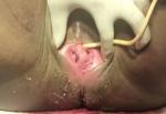
Clinical findings: she was a young woman, healthy looking, afebrile, not pale, well hydrated, nil pedal edema. Her pulse rate was 82 beats per minutes, full volume, and regular. Blood pressure was 110/70mmHg. Respiratory rate was 18 cycle per minute. Abdomen was uniformly enlarged, symphysiofundal height was 39cm, singleton fetus was palpable in longitudinal lie, cephalic presentation, fetal heart rate was 148 beats per minutes. Pelvic examination revealed two vaginal openings separated by a longitudinal septum, normal urethral and anal orifices (Figure 1)
Diagnostic assessment: the full blood count shows packed cell volume of 35%, a white blood cell count of 8 x 10⁹/L, platelet count of 250,000/ml. Blood group was O positive, HIV 1 and 2 were non-reactive, Hepatitis B surface antigen and Hepatitis C virus screening were negative. Urinalysis was essentially normal.
Diagnosis: uterine Didelphys with viable fetus at term in a multigravida with 2 previous caesarean sections.
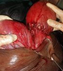
Therapeutic interventions: informed consent was obtained for elective caesarean section, and she was reviewed by the anaesthetists. She had subarachnoid block done followed by caesarean section. She was delivered electively at 37 weeks with the following intra-operative findings of Pfannenstiel scar that healed by primary intention, moderate adhesion between anterior abdominal wall and the uterus, didelphylic uterus each bearing single tube and ovary, gravid left hemi uterus. The right hemi uterus was about the size of an adult fist. A life male neonate delivered by breech, baby weighed 3.4kg with Apgar score of 8 and 9 in the 1 and 5 mins, placenta was anterior and fundal and EBL 800mls (Figure 1). She did well post operatively and was discharged home on the fifth day post-surgery after counseling for family planning.
Follow up and outcome of interventions: she was seen at the post-natal clinic 2 and 6 weeks after her discharge with no complaints and was subsequently discharged to the family planning clinic where she was placed on implant Nexplanon® for contraception.
Patient perspective: she was satisfied with the quality of care she received during her antenatal and post-natal visits.
Consent: informed consent was obtained from the patient for images and information provided in this case report.
Conception in patients with uterine didelphys is not impaired and it is considered better than patients of other Müllerian duct anomalies, however it's associated with poor pregnancy outcomes [6]. There are reports that live birth in women with uterine didelphys is less than half with about 1 in 3 pregnancies ending up in abortion, about half in premature deliveries and about 1 in 5 reaching term [3]. However others reported excellent pregnancy outcome with good management [3,6]. This patient had consecutive spontaneous conceptions, and they were carried to term. The reasons for this might be the level of development and capacity of the uterus. Many patients with this condition have no symptoms and are usually discovered during investigation for recurrent pregnancy loss or preterm labour, thus cases of uterine didelphys are largely under reported as its true incidence is not known [7]. However, some patients can present with non-classical symptoms like pelvic discomfort, dyspareunia, dysmenorrheal, hematocolpos and hematometria [8]. Our patient was asymptomatic. Diagnosis of uterine didelphys can be done with an ultrasound scan, hysterosalpingography, magnetic resonance imaging (MRI), abdominal laparoscopy and laparotomy [9]. The diagnosis of complete uterine didelphys (two uterus, two cervixes and a longitudinal vaginal septum) in our patient was made at the caesarean section for the delivery of her first pregnancy. A complete longitudinal septum is found in about 75% of cases of uterine didelphys [10]. This was also seen in our patient. Accurate diagnosis is essential to patient management, especially during pregnancy and delivery. It has been reported that about 51-80% of the pregnancies in women with uterine didelphys are delivered by caesarean section due to obstetric complications [9,10], while some other authors had also reported vaginal deliveries to the tune of 5.9% in some women with uterine didelphys [8]. Our patient had a total of three caesarean sections at term with good fetal outcome.
In conclusion, women with congenital uterine anomaly such as uterine didelphys can be diagnosed with high index of suspicion. Management of this patient can pose a serious challenge to the obstetrician, especially as pregnancy in this group of patients are associated with adverse outcomes. However, individualized management of patients with uterine didelphys and delivery by elective caesarean section produces favorable pregnancy outcome.
The authors declare no competing interests.
All the authors have read and agreed to the final manuscript.
Figure 1: two different vagina openings with its associated cervix
Figure 2: didelphys uterus with lower uterine segment incision repair on the hemi uterus following caeserean section
- Shahanaj S, Didarul A, Farzana R. Uterine Didelphys with Pregnancy Outcome: A Case Report. Chattagram Maa-O- Shishu Hospital Medical College Journal. 2018;17(2):53-55. Google Scholar
- Stanislav S, Stoyan K, Angel Y. Pregnancy and childbirth in Uterine Didelphys: A Report of Three Cases. Medicina (Kaaunas). 2020;56(4):198. PubMed | Google Scholar
- Omeed P, Dana B, Caria P, Wendy W. Uterine Didelphys in a Pregnant Mother. Open Journal of Obstetrics and Gyeacology. 2018;8(13).
- The American Fertility Society. The American Fertility Society classifications of adnexal adhesions, distal tubal occlusion, tubal occlusion secondary to tubal ligation, tubal pregnancies, m�llerian anomalies and intrauterine adhesions. Fertil Steril. 1988 Jun;49(6):944-55. PubMed | Google Scholar
- Grimbizis GF, Gordts S, Sardo ADS, Brucker S, De Angelis C, Gergolet M et al. The ESHRE/ESGE consensus on the classification of female genital tract congenital anomalies. Hum Reprod. 2013 Aug;28(8):2032-44. PubMed | Google Scholar
- Mohammad Othman. Uterine Didelphys Pregnancy Management. Journal of Advances in Medicine and Medical research. 2018;26(4);1-5. Google Scholar
- Rezai S, Bisram P, Alcantara I, Upadhyay R, Lara C, Elmadjian M. Didelphys uterus: a case report and review of the literature. Case Report in Obstetrics and Gynecology. 2015;2015:865821. PubMed | Google Scholar
- Namkha D, Sangay T, Tshering W. Uterus Didelphys with double vagina diagnosed during third caeserean section: A case report. SAGE Open Medical Case Reports. 2022 Jan 19;10:2050313X211072967. PubMed | Google Scholar
- Yadeta B, Bekele E, Kumera K, Digafe T. Pregnancy in Uterus Didelphys Delivered by Caesarean Delivery: A Case Report. GynecolObstet Case Rep. 2020;6(3):20.
- Bakari F, Adesiyun AG, Ochogwu EP, Umar-Sulayman H, Ameh N, Bawa US. Complete uterine didelphys: An incidental finding at emergency caesarean section. Arch Int Surg. 2016;6(4):233-6. Google Scholar
Search
This article authors
On Pubmed
On Google Scholar
Citation [Download]
Navigate this article
Similar articles in
Key words
Tables and figures
Article metrics

