
Nodding syndrome: an enigmatic neglected tropical disease, a newly recognized neurodegenerative disorder with regional clusters in East Africa; could it be an early-onset Alzheimer´s disease?
David Lagoro Kitara, Bernardo Lemos, Jesse Boardman Bump
Corresponding author: David Lagoro Kitara, Harvard University, Harvard T.H. Chan School of Public Health, Department of Global Health and Population, Boston, Massachusetts, United States of America 
Received: 20 May 2021 - Accepted: 24 Sep 2021 - Published: 24 Dec 2021
Domain: Tropical medicine,Neurology (general),Global health
Keywords: Nodding Syndrome (NS), Alzheimer’s disease (AD), Modified Rankin Scale (MRS), Entorhinal Cortex (EC), cognitive decline, Gulu, Uganda
©David Lagoro Kitara et al. PAMJ Clinical Medicine (ISSN: 2707-2797). This is an Open Access article distributed under the terms of the Creative Commons Attribution International 4.0 License (https://creativecommons.org/licenses/by/4.0/), which permits unrestricted use, distribution, and reproduction in any medium, provided the original work is properly cited.
Cite this article: David Lagoro Kitara et al. Nodding syndrome: an enigmatic neglected tropical disease, a newly recognized neurodegenerative disorder with regional clusters in East Africa; could it be an early-onset Alzheimer´s disease?. PAMJ Clinical Medicine. 2021;7:31. [doi: 10.11604/pamj-cm.2021.7.31.29943]
Available online at: https://www.clinical-medicine.panafrican-med-journal.com//content/article/7/31/full
Research 
Nodding syndrome: an enigmatic neglected tropical disease, a newly recognized neurodegenerative disorder with regional clusters in East Africa; could it be an early-onset Alzheimer´s disease?
Nodding syndrome: an enigmatic neglected tropical disease, a newly recognized neurodegenerative disorder with regional clusters in East Africa; could it be an early-onset Alzheimer´s disease?
![]() David Lagoro Kitara1,2,&, Bernardo Lemos3, Jesse Boardman Bump1
David Lagoro Kitara1,2,&, Bernardo Lemos3, Jesse Boardman Bump1
&Corresponding author
Introduction: Nodding Syndrome (NS) is an enigmatic childhood neurological disorder clustered in East Africa. Histo-immunopathological analysis of brains of deceased NS children showed brain atrophy, neurofibrillary tangles, and tau protein deposition mainly in the entorhinal cortex. The aim of this paper is to describe the clinical and neurological presentations, to use Modified Rankin Scale (MRS) to assess disability and observe possible similarities to an early-onset Alzheimer´s disease.
Methods: a case-control study involving 21 NS cases, 21 age and sex-matched community controls, 21 younger healthy siblings and 21 biological parents was conducted. Each NS child and controls underwent clinical and neurological examinations and MRS was used to assess the level of disability. Ethical approval was obtained, and STATA version 14.1 was used for data analysis. A p-value less than 0.05 was considered significant.
Results: children with NS exhibited significant cognitive disability in many ways, including poor immediate recall (short-term memory) 15/21(71.4%), disorientation 13/21(61.9%), muteness 4/21(19.0%), poor delayed recall 11/21(52.4%), and poor concentration 9/21(42.9%). Just over half of NS-affected children 11/21(52.4%) exhibited abnormal coordination of limb movements, but majority had normal cranial nerves 18/21(85.7%), slightly less than half had a normal gait 10/21(47.6%), and no significant association was observed between poor MRS (score ≥2) with; current age (χ2=4.039, p=0.854), underweight (χ2=1.636, p=0.201), age at onset (χ2=10.611; p=0.389), and reported duration of the syndrome (χ2=4.604, p=0.466).
Conclusion: clinical and neurological findings suggest cognitive decline is related primarily to disorders of entorhinal cortex similarly observed in early-onset Alzheimer´s disease. It may not be too early to suggest that NS is an early-onset Alzheimer´s disease.

Nodding Syndrome (NS) is an enigmatic and devastating childhood neurological disorder clustered in East Africa and characterized by pathognomonic features of rhythmic head-nodding (dorso-ventral movement), cognitive decline, behavioral disturbances, and developmental delays [1-3]. The clinical course of NS surfaces with behavioral changes and absences in previously healthy children mainly between the ages of 5-15 years at onset, which is usually followed by recurrent episodes of head-nodding spells, cognitive decline, and generalized tonic-clonic seizures at later stages [1-5]. Nodding episodes appear to be triggered by eating and other sensory stimuli such as cold weather, cold water, starvation, and febrile illnesses [1-3]. Most affected families are usually relatives and commonly have multiple children with NS compared to other community members [1-3,5].
In Uganda and Tanzania there are reportedly more than 10,000 children affected with Nodding Syndrome [1]. Currently, there is no universally agreed method for screening or identifying those at risk of developing the syndrome [5]. However, as reported in Nature´s 2018 neurological review, histo-immunopathological studies on brains of deceased NS children found brain atrophy, neurofibrillary tangles, and tau protein deposition in the entorhinal cortex of the brain, and a conclusion of tauopathy was made [6]. Macroscopically, post-mortem analyses have found that brains of NS-affected children showed mild fronto-temporal cortical atrophy, which may be linked to cognitive impairment [7] and Parkinsonism as detected in clinical presentations [8]. Similarly, neuro-pathological examination of NS brains showed tau-immunoreactive neuronal neurofibrillary tangles, pre-tangles, neuropil threads and dot-like lesions involving mainly the cerebral cortex, subcortical nuclei, and brainstem [4].
There was preferential involvement of frontal and temporal lobes in patchy distribution, mostly involving crests of gyri and superficial cortical lamina [4]. Mesencephalopontine tegmental nuclei, substantia nigra and locus coeruleus also revealed globose neurofibrillary tangles and threads [6]. Tegmentum, locus coeruleus and substantia nigra contained tangles and threads but neuronal loss and gliosis were not prominent features in the cases reported [4]. To date, there are no studies that have described the late stages of Nodding Syndrome progression to correlate the clinical and neurological features with the anticipated pathology seen in NS brains at autopsy.
Alzheimer´s disease (AD), commonly seen in the elderly over 65 years of age, presents with cognitive decline, brain atrophy, tau protein deposition, and neurofibrillary tangles [9]. Substantial tau is deposited in the entorhinal cortex of the brain in AD patients, a feature that we also observed in cases of NS. The entorhinal cortex (EC) is anatomically located in the medial temporal, frontal, parietal cerebral lobes, and functions as a hub in a widespread network for memory, navigation, and perception of time [10-12]. It is the main interface between hippocampus and neo-cortex of the cerebrum [10-12]. The EC-hippocampus system plays an important role in declarative memories and in particular spatial memories including memory formation, memory consolidation and memory optimization in sleep [10-12].
Furthermore, EC is responsible for pre-processing of input signals in reflex nictitating membrane response of classical trace conditioning and association of impulses from eyes and ears [10-12]. Therefore, entorhinal cortex is one of the most important memory centers in the brain, and it relays messages to and from hippocampus, which is viewed as one of the major sections of the brain and epicenter for long-term memory and spatial navigation [10-12].
Studies have reported that EC is one of the first areas of the brain to be affected by plaques and tangles in the build-up of Alzheimer´s disease (AD) [10-12]. From a neuro-anatomical perspective, tissues and structures of nervous system, EC holds some major responsibility in retaining neural blueprints of spatial movements [10-12]. Also, the area has a number of path cells, which helps an individual to navigate clockwise or counterclockwise paths of movements [10-12]. Tau deposition sites, brain atrophy and neurofibrillary tangles in brains of deceased NS children seem to overlap with those observed in Alzheimer´s disease.
These apparent similarities led the present authors to hypothesize that this enigmatic neurological disorder may perhaps be a new form of an early-onset Alzheimer´s disease whose triggers remain unknown and that they are so far found only clustered in children in East Africa. We reasoned that clinical and neurological examinations of NS children could help highlight possible sites of pathology in NS children and serve as a test of our hypothesis.

Study design: we conducted a community case-control study with 21 NS children, 21 younger healthy siblings, 21 biological parents of NS children, and 21 age and sex-matched community controls.
Study sites: the study was conducted in Odek at former Hope for HumaNs (HfHs) rehabilitation center in Aromowanglobo [1,2,5]. The center cares for NS children mainly from two sub counties (Odek and Awere) in Omoro and Pader districts respectively. Patients were recruited from 10 villages in (Bolo Juklebi, Lapeta, Bolo Angweng, Atede West, Paikat Akidi, Ludok, Lukee, Olam, Akoyo Lumec and Ajan), the NS epicenters in northern Uganda. One of the authors (D.L.K) is a senior physician who is familiar with the populace in northern Uganda has worked in the field of public Health and epidemiology in many NS studies, collected the data.
Recruitment strategy: we recruited a convenient sample of 21 pairs of NS children and their younger healthy siblings, 21 age and sex-matched controls who were un-related and 21 biological parents of NS children with the help of Village Health Team (VHT) leaders [1,2].
Study population: we recruited and examined NS children, healthy younger siblings, biological parents, and community controls.
Selection criteria (with inclusion and exclusion criteria): a sample of 21 confirmed NS cases between the ages of 5-21 years were selected, as defined by the 1st International scientific meeting on NS in July 2012, in Kampala, Uganda, which described inclusion criteria for NS [1,3,5].
Inclusion criteria for NS children: (1) a child confirmed in accordance with World Health Organization (WHO) case definition as probable Nodding Syndrome [1,3,5] (2) ages of 5-15 years at NS onset (3) informed consent and assent.
Exclusion criteria for NS children: (1) Has no biological parents to give information about the onset of the illness (2) children less than 3 years and those with reported history of abnormal physical, cognitive, and social development prior to onset of nodding (3) lack of informed consent from parents (4) children who were acutely ill with fever during the study period.
Community controls
Inclusion criteria: (1) Healthy, age and sex-matched with NS child (2) informed consent and assent (3) no evidence of neurological disorder (4) normal childhood development as described by parents (5) no biological relationship with the NS Child.
Exclusion criteria: (1) a relative of the NS child.
Younger healthy NS siblings
Inclusion criteria: (1) no features of neurological abnormalities (2) informed consent and assent (3) three years of age or older.
Exclusion criteria: (1) did not live with the family during the IDP period.
Parents of NS children
Inclusion criteria: (1) confirmed as biological parent of NS child (2) informed consent.
Exclusion criteria: (1) contested parenthood of a NS child.
Study instruments: we used a health questionnaire, clinical and neurological examination forms, and Modified Rankin Scale [13-16].
Data collection: we collected data from each participant using a face-to-face questionnaire interview which lasted between 45-60 minutes at the former HfH rehabilitation center. These included the socio-demographic characteristics of participants including age, sex, highest level of education, occupation, addresses, religion, age at onset of the syndrome, duration of treatment and rehabilitation, nodding episodes whether observed or reported and a series of questions on medical and family history, and the clinical signs and symptoms of NS (Annex 1). This was accompanied by a complete clinical and neurological examinations which included assessment of the mental health status, short-term memory, level of concentration, the twelve cranial nerves, delayed recall at 5 minutes, the evaluation of gait, muscle strength, sensory nerve assessment, tests on reflexes and coordination of limb movements (Annex 2), and used Modified Rankin Scale (MRS) [13-16] to grade the level of neurological disability (Annex 3).
Ethical approval: this study was approved by a local IRB (Lacor Hospital, LHIREC No. 097/5/19). Written informed consent and assent were obtained from each participant. The informed consent form had the purpose, objectives, risks, and benefits of the study to participants. Also, participants were de-identified and assured on confidentiality of information, security of data and how it would be maintained. In the same way, the study was conducted in accordance with good clinical practice.
Data management: all data were double-checked and entered in Excel software and later exported to STATA version 14.1 for cleaning and analysis [17].
Data analysis: a statistical package STATA version 14.1 was used for data analysis [17], a p-value less than 0.05 was considered statistically significant. Descriptive statistics were used on participants´ socio-demographic characteristics, medical and family history where mean, median, mode, standard deviations (SD), variance, skewness, and kurtosis were calculated. To establish factors associated with NS, bivariate analyses were conducted using chi square tests (χ2). In further analysis on NS children, we used MRS as the outcome variable and compared the difference between MRS scores (≥2) and others (MRS ≤2). Notably, variables that were used in the analyses were premised on results obtained from earlier studies conducted on Nodding Syndrome in Uganda such as age at onset, current age, length of the syndrome and when it was first noted [1-3,5,7].
From May to July 2019, we conducted this case-control study at the NS epicenter in northern Uganda. Figure 1 is a bar graph presenting Modified Rankin scale (MRS), current age, age at NS onset, reported duration of the syndrome and duration of treatment of NS children. As seen, there were no marked variations among MRS, current age, age at onset and reported duration of the syndrome and treatment. Table 1 is a descriptive statistic of the study participants which showed that majority of NS children had lower anthropometric measurements compared to their age and sex-matched community controls. NS children were 82.29%, 96.85% and 87.28% of the mean weight, height and BMI of their age and sex-matched community controls respectively.
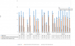
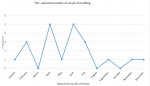
In Table 2, the chi square (χ2) tests showed that all variables on NS children were significantly different from the community controls except, presence of jaundice, history of epileptic fits and underweight. Similar observations were made with the younger healthy siblings. As we explored the onset of the syndrome, months and years of nodding onset were noted. In Figure 2, most respondents reported April and June as months for nodding onset and in Figure 3, the line graph showed that 12 years was the peak duration of the syndrome and 5 years, peak duration for the treatment and rehabilitation at the former HfH rehabilitation center.
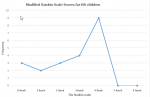
After conducting systematic clinical and neurological examinations on all study participants, we found that most NS children had Modified Rankin scale (MRS) scores of four, an indication that they had advanced disability (Figure 4). Poor Modified Rankin Scale score (MRS) (≥2) in Table 3, showed no significant association with current age, BMI, age at onset, and reported duration of the syndrome. Similarly, clinical, and neurological findings summarized in Table 4 found the most significant clinical and neurological observation was cognitive decline. Most NS children were disoriented 13/21(61.9%); mute 4/21(19.0%); had poor immediate recall 15/21(71.4%); poor delayed recall 11/21(52.4%); normal cranial nerves 18/21(85.7%); normal gait 10/21(47.6%); abnormal coordination of limb movements 11/21(52.4%); normal reflexes 17/21(81.0%); normal sensory function to crude touch and pain 14/21(66.7%); normal motor functions 16/21(76.2%), and poor concentration 9/21(42.9%).
Of special interest were the minority 3/21(14.3%) who presented with diplopia, ptosis, impaired adduction, upward and downward gaze of eyes, an indication of oculomotor nerve dysfunction; 8/21(38.1%) were ataxic, 7/21(33.3%) had sensory stimuli deficiency to crude touch and pain, 1/21(4.8%) with hemiparesis and 2/21 (9.2%) with quadriparesis.
Funding: this project was conducted with the support of the Takemi Program in International Health at Harvard T.H. Chan School of Public Health and additional support from individual authors.
Ethical approval: this study was approved by a local IRB (Lacor Hospital IREC) LHIREC No. 097/5/19 and Harvard University, T.H Chan School of Public Health. Each participant gave a written informed consent to participate and consented to have this information published.
This study was conducted in an area of northern Uganda where Nodding Syndrome is relatively common. The most significant finding was cognitive decline and poor Modified Rankin Scale (MRS) scores for majority of NS participants. Poor neurological function and disability observed in NS children have been the most observed evidence of progression of the syndrome where neurological disability becomes more advanced and physical functionality progressively reduced over time (Figure 1). Histo-immunopathological analysis of brains of deceased NS children from the same area in northern Uganda showed brain atrophy, tau protein and neurofibrillary tangle deposition in the entorhinal cortex of the brain [4].
Anthropometric measurements showed most NS children in the current study were underweight and stunted (Table 1). Whether the underweight and stunting occurred prior to the onset of the syndrome or were effects of NS remained undetermined. Owing to the long interval between NS on-set and the current study, we were unable to pin-point the actual timing of the initial occurrence of the under-nutrition. However, our long observation of NS children at HfH center from 2012 to 2018 and reports from their parents showed that their nutritional status declined more observably after the onset of the syndrome and has since followed a downward turn. The nutritional status of most NS children who were at the HfH rehabilitation center however improved remarkably with evident weight and height gains in a few months [1,3,5]. This catch-up weight and height gains were attributable to the good care and food rehabilitation at the center which presented favorable environment compared to homes of NS children where there was often food shortage [1,3,5] (Figure 1).
In spite the good physical changes seen on NS children as evident by weight and height gains while at the HfH rehabilitation center, NS children remained confronted with mental, emotional, neurological, and cognitive challenges. A formal study to review the mental and emotional challenges faced by NS children is warranted to understand the extent and gravity of the syndrome. Most clinical features on NS children compared to their community controls at bivariate analysis were significantly different except; the presence of jaundice, history of epileptic fits, and underweight (Table 2). Similarly, younger healthy siblings had comparable observations at bivariate analysis except; the number of younger healthy siblings with underweight were more compared to NS children (Table 2). Underweight in NS children and younger healthy siblings at almost equal numbers drew the attention of authors to question whether there may be some household factors that were responsible for this observation (Under-nutrition). Authors have asked questions whether there could be genetic, environmental, nutritional, medical, and socio-economic factors in NS families and homes that were responsible for this occurrence. Owing to the small sample size of this study population and limited longitudinal measurements, authors cautiously considered that further exploration of this finding in the future would be required.
It was equally important to note that the reported months of nodding onset for majority of NS children was April and June which coincided with dry spells, food shortage and food insecurity at the epicenter (Figure 2). The reported months of nodding onset in NS children was consistent with earlier research findings on NS in Uganda [1-3,5,7]. The reported duration of NS, the period of treatment and rehabilitation at the HfH Center peaked at 12 and 5 years respectively (Figure 3). This finding was consistent with previously documented results on the onset and progression of Nodding Syndrome in northern Uganda [1-3,5,7] where most cases of NS began and were discovered during the Internally Displaced persons (IDP) camps when food was scarce, and camps congested with high prevalence of infectious diseases among IDP residents.
Similarly, MRS scores for majority of NS children in this study population was four (Figure 4) and this describes the disability of a person as, “moderate to severe disability where the person is unable to attend to own bodily needs without assistance, and unable to walk unassisted” [13-16]. This finding suggests that most NS children had developed advanced neurological disease; the level of disability was high and physical functionality low. Also, poor MRS in this study population was not dependent on; age of NS onset, current age, reported duration of treatment and duration of the syndrome (Figure 1 and Table 3). These authors were unable to give explicit explanation to this observation though it was agreed that perhaps the occurrence of Nodding Syndrome as a spectrum may in part explain the variation in the level of disability, response to treatment and rehabilitation of individual NS child at the HfH Center. Secondly, authors considered the role of anti-seizure medications, food supplementation and use of multivitamins during rehabilitation at the HfH center in altering the outcome and improving the quality of life of NS children. Lastly, the nature of this study (Case-control) and the limited follow-up data on NS children in a longitudinal study may in part explain our inability to give explicit explanations on these observations so far made.
The study on mental and neurological status of NS children showed the severity of disabilities. The most significantly observed disability among NS children in the current study was cognitive decline. The majority were disoriented and mute (80.9%), had poor immediate recall (short-term memory) (71.4%), poor concentration (42.9%), and poor delayed recall (52.4%) (Table 4). This finding is supported by a United States Center for Disease Control and Prevention (CDC) led study where NS children were found to have brain atrophy on Magnetic Resonance Imaging (MRI), gross cognitive impairment, school drop-out, frequent attacks of nodding and convulsions which did not permit them to properly comprehend information [7]. The CDC study further noted that NS children were worse on cognitive tasks than their age-matched controls, with significantly lower scores on tests of short-term recall and attention, semantic fluency, and fund of knowledge, as similarly observed in this current study population [7].
In addition, minority neurological findings have attracted the interest of authors where the most affected cranial nerve in the study population was the oculomotor nerve (Cranial nerve III). The signs of oculomotor nerve dysfunction observed were diplopia, ptosis, impaired adduction, upward and downward gaze of the eyes. These oculomotor dysfunction symptoms may have resulted from either lesion of the cerebellar flocculus causing dysfunction of the retinal image stabilization or from the dorsal vermis (VI and VII) and the fastigial nuclei, resulting in saccadic dysmetria. This finding is supported by the report from a histo-immunopathological study conducted on NS brains which showed there were cerebellar degeneration [4]. Again, in Table 4, we argue that the abnormal coordination of limbs observed in NS children were likely related to cerebellar dysfunction because some NS patients presented with hypotonia, hyporeflexia, delayed onset and offset, and slowing of voluntary movements, ataxia, and tremors. Furthermore, the postural ataxia observed in some NS children may also be a pointer to cerebellar dysfunction. It is observed that postural ataxia is a condition that commonly results from lesions of the anterior lobe, the vermal parts of the vestibular cerebellum and dysfunction of cerebellar afferents.
Also, the long latency response seen in NS children which was significantly prolonged could perhaps be a result of the anterior cerebellar lobe atrophy. In addition, the abnormal coordination of limb movements reported in Table 4 were unlikely to be sensory ataxia as the ataxia we observed were minimal when NS patients were seeing their movements but worst when their eyes were closed. Furthermore, 7 out of 21 NS children we reported with sensory loss had exhibited lack of sensation and quick withdrawal reactions to crude touch and pain stimuli.
Histological descriptions of brains of deceased NS children from the same area in northern Uganda showed tau deposition in frontal cortex in gyral crowns, neurofibrillary tangles, and dystrophic neuritis in the cerebrum and brain stem [4]. The anatomical sites which were clinically most affected in NS children were mainly in the entorhinal cortex of the brain where cognitive functions are coordinated and controlled [10-12]. The most significantly observed neurological dysfunctions in most NS children were memory loss, muteness, and disorientation (Table 4). These clinical symptoms mentioned above are functions that are coordinated and controlled in the entorhinal cortex of the brain. The clinical findings on NS children overlap with those observed in Alzheimer's disease (AD), a progressive neurological disease where dementia symptoms gradually worsen over several years [10-12,18]. However, in its early stages, memory loss is usually mild, but in late-stage Alzheimer's disease, individuals lose their ability to carry on conversation and failure to respond to the environment [10-12,18]. The clinical signs observed in majority of NS children were disorientation, muteness, and loss of memory (poor short-term memory, poor concentration, and poor delayed recall) and in some, could not respond to the environment (Table 4).
Alzheimer´s disease (AD) and Nodding Syndrome (NS)
It has been observed that when AD occurs, amyloid plaque accumulation occurs in the brains of the affected person [10-12,18]. Amyloid plaque is a type of fibrous protein build-up which is considered the biological hallmark of Alzheimer's disease [10-12,18]. Once plaques collect in the neo-cortex, they affect the entorhinal cortex, making it one of the earliest affected areas of Alzheimer's-affected brains [10-12,18]. Similarly, neurofibrillary tangles build up in the entorhinal cortex and afterwards extent to other parts of the brain [10-12,18]. These pathological findings were too observed in brains of deceased NS children in the histo-immunopathological studies we reported [4].
Although plaques and tangles are pathologies that can occur in the brain, the exact mechanism on how they cause cognitive decline in AD remains unclear. The accrual of these substances (plaques, tangles, and tau protein) is thought to directly impact on the entorhinal cortex's ability to function properly [10-12,18]. As observed in many cases of AD, the entire area atrophies during AD however, the cortical atrophy observed in NS appeared mild, perhaps a milder form of the disease or may be a disease in progress [4]. How plaques and tangle deposition in the entorhinal cortex of NS brains cause the pathological presentations observed are not well understood but it is thought it contributes to the pathogenesis of NS in the same way as in Alzheimer's disease.
These pathological processes (brain atrophy, tangles and plagues formation and deposition) in the entorhinal cortex of the brain are thought to result in major problems of short-term memory preservation, memory consolidation and spatial navigation [9,18] which were observed in most NS children in the current study population (Table 4). Nevertheless, the absence of neuritic plaques and limbic tau pathology in NS brains uniquely demonstrates the pathological difference between AD and NS. The sparing of the hippocampus (there are no tau and neurofibrillary tangle deposition) in NS cases we examined marks one of the major differences between Nodding Syndrome and Alzheimer´s disease. However, considering that we had limited number of samples examined, it may be too early to rule out other pathologies which were not observed in these NS cases.
Nodding Syndrome (NS) and other neurodegenerative disorders (ND)
Many chronic and progressive neurodegenerative disorders are characterized by selective and symmetric loss of neurons in motor, sensory and cognitive systems [9]. Delineation of the pattern of cell loss and identification of disease-specific cellular markers have helped in nosological classification of neurological disorders. For example, senile plaques, neurofibrillary tangles, neuronal loss, and acetylcholine deficiency defines Alzheimer´s disease [9,11,12]; Lewy bodies and depletion of dopamine characterizes, Parkinson´s disease [9,19]; cellular inclusions and swollen motor axons are found in amyotrophic lateral sclerosis [20]; and γ-aminobutyric acid-containing neurons of the neostriatum are lost in Huntington´s disease [21]. In addition, Mendelian inheritance are demonstrable in many neurological disorders [9] for example in Huntington´s disease, a family history of the disease can be ascertained in most cases [22]. Similarly, clinical, and epidemiological observations from studies conducted in Uganda found that most NS children were clustered in specific families and in relatives [1,3,5,23].
Also, 1% to 10% of all neurodegenerative disorders are inherited, often as autosomal dominant traits for example in Alzheimer´s disease [11], Parkinson´s disease [12] and amyotrophic lateral sclerosis [20]. Findings in this current study suggest that additional research is warranted to ascertain whether NS is inherited as other neurodegenerative disorders described above. It is suggested that comprehensive genetic and epigenetic studies on NS clusters should be conducted, and that East Africa would be the preferred site to begin with.
The role of Tau in formation of neurofibrillary tangles
Some researchers have considered the formation of paired helical filaments and neurofibrillary tangles by tau molecules as the primary pathogenic mechanism in Alzheimer´s disease [12,24,25]. This has been bolstered by a recent discovery of mutations in tau that are associated with neurofibrillary tangles and dementia [12,24,25]. Neurofibrillary tangles and tau protein deposition in the entorhinal cortex of the brain provide the resemblance between nodding syndrome and Alzheimer´s disease [4]. There are additional similarities between Nodding Syndrome and Alzheimer´s disease in that both are progressive neurological diseases which result in irreversible loss of neurons, particularly in the entorhinal cortex [4,26]. The clinical hallmarks of NS as observed in the current study is impairment of memory, judgment, decision making, orientation to physical surroundings and language (Table 4). In addition, the CDC led study on NS in Uganda found progressive cognitive decline in NS children compared to their age and sex-matched community controls. Also, the provisional diagnosis of AD is usually based on clinical and neurological examinations, with exclusion of other causes of dementia and, a definitive diagnosis is made at autopsy [12,24,25]. This observation suggests that clinical and neurological examinations have a role in describing Alzheimer´s disease as well as other neurological disorders and perhaps nodding syndrome inclusive.
Potential limitations of this study and how we addressed them
This community case-control study was conducted in two months in 2019. Its limitations are based on the study design and a small sample population. A larger sample size of research participants would have produced a much more powered results for the observations and outcomes we have reported. In addition, longitudinal measurements of variables especially neurological status and Modified Rankin Scale (MRS) would have helped determine the progressiveness of the cognitive decline. However, considering that NS children were born normal, had normal childhood milestones, physical, cognitive, emotional, and behavioral developments just as their age and sex-matched community controls before nodding onset, we can reasonably suggest that there has been a progressive cognitive decline due to the syndrome. In addition, we conducted detailed clinical analysis, physical and neurological examinations and used Modified Rankin Scale (MRS) to study and score each study participant. This, we considered was in-depth enough to provide generalizable information which were useful.
This study had a problem of selecting NS children who were present at the time of the study and were willing to be interviewed and examined. This may have created some selection bias of participants. However, this also allowed us to select only probable and confirmed NS cases and thus permitted us to obtain all the required information from participants with no missing data. The clinical characteristics of NS children were quite typical compared to other neurological diseases and we were able to identify only probable and confirmed cases of NS to compare with the age and sex-matched community controls. Recall and social desirability biases from the interviewer-administered questionnaire were also potential limitations to this study. For example, parents of NS children may have hesitated to inform interviewers on other life bothering situations that could be affecting them for example stress because of personal pressures, undesirable family issues, divorce, domestic violence, deaths, and stigma resulting from NS sickness in their families. These social issues were not explored in detail but could form a basis for further studies on NS children and their families.
There could have been more confounders that may have been residual that could have arisen due to misclassifications of measured or reported variables on NS children. These could include factors such as fatigue, current medications (anticonvulsants-sodium valproate) which affect mentation of NS children and other liabilities which we could not determine. Similarly, we were unable to determine and eliminate all confounders, but we hope that in future we should conduct more comprehensive prospective cohort studies and have multiple longitudinal measurements of variables on NS children to determine the progression of this syndrome.
We acknowledge that the tau deposition in the 5 deceased NS brains were negative for amyloid beta type as described in the modern definition of Alzheimer´s disease. As numbers of cases studied were our limitation and as more studies are being conducted, it may be premature to conclusively rule out additional findings on the characteristics of the tau deposition in NS children.
Generalizability of the information obtained: the information generated from this study can be generalizable to NS patients in Northern Uganda and particularly NS patients that were cared for in a well-established rehabilitation center for at least a few years.
In conclusion, the clinical findings in NS children suggest that there is cognitive decline. This neurological disability is associated with disorders of the entorhinal cortex which are similarly observed in an early-onset Alzheimer´s disease. Perhaps, it may not be too early to suggest that NS is reminiscent to an early-onset Alzheimer´s disease. These authors, however, recommend more comprehensive studies on epigenetics and genetics of NS including characterization of tau proteins in brains of NS children.
What is known about this topic
- Nodding syndrome is one of the childhood neurological disorders of unknown cause;
- It is clustered in East Africa (Uganda, Tanzania, and South Sudan);
- Nodding episodes are triggered by eating food, cold weather, febrile illnesses, and starvation.
What this study adds
- The main pathology in NS children at autopsy are brain atrophy, tau, and neurofibrillary tangle deposition in the entorhinal cortex of the brain;
- Clinical and neurological examinations of children with nodding syndrome revealed severe neurological and cognitive disabilities. The most affected functions are those coordinated and controlled at the entorhinal cortex of the brain;
- The cognitive decline is progressive and shows similarity to progression in an early-onset Alzheimer´s disease.
The authors declare no competing interests.
DLK contributed to the conceptualization, literature review, study design, data collection and analysis, interpretation and write up of the manuscript; BL and JBB analyzed the data, made critical revision, and wrote the manuscript. All authors have read and agreed to the final manuscript.
We wish to acknowledge with many thanks the generous support from staff of Omoro district local government managing the former Hope for HumaNs rehabilitation center (HfH) in Odek Sub County for the care and space for examining Nodding Syndrome children. We deeply acknowledge our research teams for data well collected (Dr. Tabu Kenneth, Dr. Mwaka Joseph, Dr. Akello Cissy, Dr. Oceng Denis Oloya, Dr. Orech Daniel, Dr. Oryema Bosco and Dr. Opupe Richard). We further acknowledge Guo MuQi for the well conducted statistical analysis. Our most appreciation to Prof. Michael Pollanen and his team for the histological slides.
Table 1: descriptive statistics of the case-control study
Table 2: bivariate analysis of NS cases with matched community controls and younger siblings
Table 3: factors associated with poor MRS (≥2) among NS children
Table 4: summary of the neurological and mental health status of the 21 NS children
Figure 1: current age, reported duration of the syndrome and rehabilitation, and Modified Rankin Scale (MRS) of NS participants
Figure 2: reported months of nodding onset among nodding syndrome children
Figure 3: reported duration of the syndrome and duration of intervention on nodding syndrome participants
Figure 4: Modified Rankin Scale (MRS) for nodding syndrome participants
Annex 1: health questionnaire
Annex 2: neurological examination form
Annex 3: Modified Rankin Scale (MRS) for NS participants
- Anywar Arony Denis, Angwech Collins, Makumbi Edward Frederick, Suzanne K Gazda, Kitara David Lagoro. Is there a line between internal displacement, environmental and Dietary factors in the onset of nodding syndrome in northern Uganda? A clinical observational study design. World J Pharma and Med Res. 2017;3(9):34-48.
- Landis Jessa L, Palmer Valerie Spencer, Spencer Peter S. Nodding syndrome in Kitgum District, Uganda: association with conflict and internal displacement. BMJ Open. 2014 Nov 4;4(11):e006195. PubMed | Google Scholar
- Kitara David Lagoro, Anywar AronyDenis, Mwaka Amos Deogratius, Uwonda Gilbert, Abwang Bernard, Kigonya Edward. Nodding syndrome in Northern Uganda: a probable metabolic disorder. Br J Med Med Res. 2013;3(4):2054-2068. Google Scholar
- Michael Pollanen S, Sylvester Onzivua, Janice Robertson, Paul McKeever M, David Lagoro Kitara, Amy Wong. Nodding syndrome in Uganda is a tauopathy. Acta Neuropathologica. 2018;136(5):691-697. Google Scholar
- Spencer Peter S, Kitara David Lagoro, Gazda Suzanne K, Winkler Andrea S. Nodding syndrome: 2015 international conference report and Gulu Accord. eNeurologicalSci. 2016;3:80-83. Google Scholar
- Charlotte Ridler. Nodding syndrome discovered as a Tauopathy. Nature reviews Neurology. 2018 Nov;14(11):632. PubMed
- Sejvar James J, Kakooza AM, Foltz JL, Makumbi I, Atai-Omoruto AD, Malimbo M et al. Clinical, neurological, and electrophysiological features of nodding syndrome in Kitgum, Uganda: an observational case series. Lancet Neurol. 2013 Feb;12(2):166-74. PubMed | Google Scholar
- Spencer Peter S, Palmer Valerie Spencer, Jilek-Aall Louise. Nodding syndrome: origins and natural history of a longstanding epileptic disorder in sub-Saharan Africa. Afr Health Sci. 2013 Jun;13(2):176-82. PubMed | Google Scholar
- Joseph Martin B, Franklin Epstein H. Mechanisms of disease; molecular basis of neurodegenerative disorders. The New Engl J Med. 1999;340(25):1970-1980. PubMed | Google Scholar
- Hyman BT, Van Hoesen GW, Damasio AR. Memory-related neural systems in Alzheimer´s disease: an anatomic study. Neurol. 1990 Nov;40(11):1721-30. PubMed | Google Scholar
- Selkoe DJ. Molecular pathology of Alzheimer´s disease: the role of amyloid. In: Growdon JH, Rossor MN, eds. The dementias. Vol 19 of Blue books of practical neurology. Boston. Butterworth-Heinemann. 1998.
- Clark CM, Ewbank D, Lee VMY, Trojanowski JQ. Molecular pathology of Alzheimer´s disease: neuronal cytoskeletal abnormalities. In: Growdon JH, Rossor MN, eds. The dementias. Vol. 19 of Blue books of practical neurology. 1998. Boston. Butterworth-Heinemann.
- Patel N, Rao VA, Heilman-Espinoza ER, Lai R, Quesada RA, Flint AC. Simple and reliable determination of the modified rankin scale in neurosurgical and neurological patients: the mRS-9Q. Neurosurgery. 2012;71(5):971-5. PubMed | Google Scholar
- Bruno A, Shah N, Lin C. Improving modified rankin scale assessment with a simplified questionnaire. Stroke. 2010;41(5):1048-50. PubMed | Google Scholar
- Quinn TJ, Dawson J, Walters M. Dr John Rankin; his life, legacy, and the 50th anniversary of the rankin stroke scale. Scott Med J. 2008;53(1):44-7. PubMed | Google Scholar
- Wilson JL, Hareendran A, Hendry A. Reliability of the modified rankin scale across multiple raters: benefits of a structured interview. Stroke. 2005;36(4):777-781. PubMed | Google Scholar
- StataCorp. Stata statistical software: release 14. college station, TX: statacorp LP. 2015.
- Cummings JL, Vinters HV, Cole GM, Khachaturian ZS. Alzheimer´s disease: etiologies, pathophysiology, cognitive reserve, and treatment opportunities. Neurol.1998;51:Suppl;1:S2-S17. PubMed | Google Scholar
- Edwards RH. Molecular analysis of Parkinson´s disease. In: Martin JB, ed. Molecular neurology. New York. Scientific American. 1998
- Brown RH. Amyotrophic lateral sclerosis and the inherited motor neuron diseases. In: Martin JB, ed. Molecular neurology. New York. Scientific American. 1998.
- Kowall NW, Ferrante RJ, Martin JB. Patterns of cell loss in Huntington´s disease. Trends Neurosci. 1987;10(1):24-9. Google Scholar
- Young AB. Huntington´s disease and other trinucleotide repeat disorders. In: Martin JB, ed. Molecular neurology. New York. Scientific American. 1998.
- David Lagoro Kitara, Jason Oh, Mwaka Amos Deogratius. Nodding Syndrome in Uganda-a disease cluster: An epidemiological dilemma. Pacific J Med Sci. 2013;11(1):21-33. Google Scholar
- Hardy J, Gwinn-Hardy K. Genetic classification of primary neurodegenerative disease. Science. 1998 Nov 6;282(5391):1075-9. PubMed | Google Scholar
- Goedert M, Spillantini MG, Davies SW. Filamentous nerve cell inclusions in neurodegenerative diseases. Curr Opin Neurobiol. 1998;8(5):619-32. Google Scholar
- McKhann G, Drachman D, Folstein M, Katzman R, Price D, Stadlan EM. Clinical diagnosis of Alzheimer's disease: report of the NINCDS-ADRDA Work Group under the auspices of Department of Health and Human Services Task Force on Alzheimer's disease. Neurol. 1984 Jul;34(7):939-44. PubMed
Search
This article authors
On Pubmed
On Google Scholar
Citation [Download]
Navigate this article
Similar articles in
Key words
Tables and figures
Article metrics

