
The Tornwaldt cyst
Zakaria Toufga, Meriem Fikri
Corresponding author: Zakaria Toufga, Department of Neuroradiology, Speciality Hospital, Rabat, Morocco 
Received: 14 Nov 2019 - Accepted: 15 Nov 2019 - Published: 25 Nov 2019
Domain: Otolaryngology (ENT)
Keywords: Tornwaldt cyst, nasopharynx, MRI
©Zakaria Toufga et al. PAMJ Clinical Medicine (ISSN: 2707-2797). This is an Open Access article distributed under the terms of the Creative Commons Attribution International 4.0 License (https://creativecommons.org/licenses/by/4.0/), which permits unrestricted use, distribution, and reproduction in any medium, provided the original work is properly cited.
Cite this article: Zakaria Toufga et al. The Tornwaldt cyst. PAMJ Clinical Medicine. 2019;1:26. [doi: 10.11604/pamj-cm.2019.1.26.20984]
Available online at: https://www.clinical-medicine.panafrican-med-journal.com//content/article/1/26/full
Case report 

The Tornwaldt cyst
The tornwaldt cyst
Zakaria Toufga1,&, Meriem Fikri1
1Department of Neuroradiology, Speciality Hospital, Rabat, Morocco
&Corresponding author
Zakaria Toufga, Department of Neuroradiology, Speciality Hospital, Rabat, Morocco
We report the case of a patient who presented otological and neurological symptoms; an endoscopic examination has objectified formation of the nasopharynx and the imaging has confirmed the cystic nature of this formation compatible with a Tornwaldt cyst. This lesion is quite rare, benign and often asymptomatic, its knowledge is important to eliminate other malignant causes and invasive management.

The Tornwaldt cyst is a benign cystic lesion that develops in the posterior medial wall of the nasopharynx, the etiopathogeny is still controversial, several clinical signs can guide the diagnosis but the discovery is in the majority of fortuitous cases and the MRI represents the examination of choice.

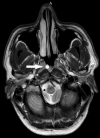
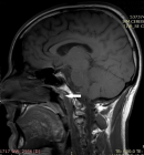
A 32-year-old woman admitted for a sensation of fullness and buzzing in her right ear with repeated episodes of otitis. The blood test was normal. Endoscopy revealed a solitary nodule in the posterior wall of the nasopharynx. A computed tomodensitometry (CT) showed the presence of a nodular formation of the posterior wall of the nasopharynx, lateralized on the right with a fluid density and without enhancement after injection of contrast medium; magnetic resonance imaging (MRI) confirmed the fluid nature of the lesion by the T2 hyper signal (Figure 1) and the T1 intermediate signal (Figure 2) allowing the diagnosis of a Tornwaldt cyst.
The Tornwaldt cyst is a benign cystic lesion that develops in the posterior medial wall of the nasopharynx; its incidence is 4% on autopsy samples, however an incidence of 0.2 to 5% has been observed on routine brain and cervical MRI [1,2]. The etiopathogeny remains controversial, some assume that it is a relic of the notochord (embryonic tissues from which vertebrae are formed), while others evoke an iatrogenic occlusion of normal structure after adenoidectomy or chronic inflammation [3]. The Tornwaldt cyst is most often asymptomatic; however, an increase in its volume or inflammation may cause occipital headache, nasal obstruction with persistent discharge, bad breath, cervical myalgia, eustachian tube dysfunction, and sometimes infections of the middle ear [3]. During endoscopic examination, the cyst appears as a solitary, medial and superficial mass on the posterior medial wall of the nasopharynx; its size can vary from a few millimeters to two centimeters with sometimes visualization of a drainage hole [1]. CT visualizes the largest cysts in the form of a fluid density lesion, but MRI remains the reference examination to identify this lesion, with a hypo or a T1 signal (depending on the protein content of the cyst) and a hyper signal T2 [1,4]. The main differential diagnoses are: Rathke's pocket cyst, meningocele, meningoencephalocele and necrotic nasopharyngeal tumors [1]. Surgical treatment is reserved for symptomatic forms outside a possible infectious episode, either by nasal endoscopy, or by oral retro palate [3].
The Tornwaldt cyst is a congenital cystic formation developed in the posterior part of the nasopharynx; it is a rare benign lesion whose diagnosis must be known to eliminate any malignant cause and eventual invasive treatment.
The authors declare no competing interest.
All the authors have read and agreed to the final manuscript.
Figure 1: T2-weighted axial section showing a well-defined cystic lesion, lateralized to the right at the posterior wall of the nasopharynx
Figure 2: T1-weighted sagittal section showing cystic lesion of posterior wall of nasopharynx in iso-signal
- Magliulo G, Fusconi M, D'Amico R, de Vincentiis M. Tornwaldt's cyst and magnetic resonance imaging. Ann Otol Rhinol Laryngol. 2001;110(9):895-6. PubMed | Google Scholar
- Miyahara H, Matsunaga T. Tornwaldt's disease. Acta Otolaryngol Suppl. 1994;517:36-9. PubMed | Google Scholar
- Maliyappanahalli Siddappa Vijayashree BV. Tornwaldt´s Cyst, Tornwaldt´s Bursitis and Tornwaldt´s Disease: a review. Research in Otolaryngology. 2014;3(4):60-63. Google Scholar
- Ikushima I, Korogi Y, Makita O, Komohara Y, Kawano H, Yamura Y et al. MR imaging of Tornwaldt's cysts. AJR Am J Roentgenol. 1999;172(6):1663-5. Google Scholar
Search
This article authors
On Pubmed
On Google Scholar
Citation [Download]
Navigate this article
Similar articles in
Key words
Tables and figures
Article metrics
PlumX Metrics
The Tornwaldt cyst
