
Outcomes of derotation osteotomy at synostosis site for congenital radioulnar synostosis
Tazi Charki Mohammed, Abdellaoui Hicham, Atarraf Karima, Chater Lamyae, Afifi Moulay-Abderahman
Corresponding author: Tazi Charki Mohammed, Department of Orthopaedic Paediatric, University Sidi Mohammed Benabdellah, University Hospital Hassan II, Fez, Morocco 
Received: 21 Oct 2019 - Accepted: 05 Nov 2019 - Published: 29 Nov 2019
Domain: Orthopedic surgery,Pediatric surgery
Keywords: Radioulnar synostosis, hyperpronation deformity, derotation osteotomy, synostosis site, complications
©Tazi Charki Mohammed et al. PAMJ Clinical Medicine (ISSN: 2707-2797). This is an Open Access article distributed under the terms of the Creative Commons Attribution International 4.0 License (https://creativecommons.org/licenses/by/4.0/), which permits unrestricted use, distribution, and reproduction in any medium, provided the original work is properly cited.
Cite this article: Tazi Charki Mohammed et al. Outcomes of derotation osteotomy at synostosis site for congenital radioulnar synostosis. PAMJ Clinical Medicine. 2019;1:31. [doi: 10.11604/pamj-cm.2019.1.31.20680]
Available online at: https://www.clinical-medicine.panafrican-med-journal.com//content/article/1/31/full
Case series 

Outcomes of derotation osteotomy at synostosis site for congenital radioulnar synostosis
Outcomes of derotation osteotomy at synostosis site for congenital radioulnar synostosis
Tazi Charki Mohammed1,&, Abdellaoui Hicham1, Atarraf Karima1, Chater Lamyae1, Afifi Moulay-Abderahman1
1Department of Orthopaedic Paediatric, University Sidi Mohammed Benabdellah, University Hospital Hassan II, Fez, Morocco
&Corresponding author
Tazi Charki Mohammed, Department of Orthopaedic Paediatric, University Sidi Mohammed Benabdellah, University Hospital Hassan II, Fez, Morocco
Congenital radioulnar synostosis (CRUS) is rare upper limb malformation in which proximal portions of the radius and ulna fuse with each other and restricts the rotation of the forearm. In cases with severe deformity, performing activities of daily living is difficult, and a surgical intervention is recommended. We present a retrospective study including patient operated for CRUS between January 2009 and December 2017 by derotation in synostosis site. Ten patients were operated. Five was unilateral and five bilateral (only one patient was operated for both sides. Mean age was 5 years. Preoperative assessment revealed limitation of the activities of daily life in all patients with an average pronation deformity of 62o. Type III of Cleary and Omer classification was the most frequent. The average correction of the forearm angle was 54o. Nine patients were satisfied and showed improvements in the ability to perform the activity of daily life. We report one case of complication (compartmental syndrome with nonunion). In summary, CRUS is a rare deformity, frequently bilateral and more commonly seen in male patients. Indications for surgery are pronation deformity of 60o or more, bilaterality and pronation of 20o to 60o with disability. It has been reported that complications are more common in osteotomy in synostosis site.

Congenital radioulnar synostosis (CRUS) is a rare upper limb malformation [1]. Since the first description by Sandifort in 1793 [2], only 350 cases have been reported in literature heretofore [3]. It is thought to be caused by a failure of longitudinal segmentation with persistence of the cartilaginous anlage between the radius and ulna during the seventh week of development. The resultant bridge may be fibrous or bony [4,5]. CRUS may be isolated or associated with other abnormalities such us brachydactyly, polydactyly, syndactyly and Madelung's deformity and can be a part of syndromes (arthrogryposis, Apert´s syndrome, Carpenter´s syndrome, Williams' syndrome) [6]. The patients with CRUS have a fixed forearm in positions that vary from neutral rotation to considerable pronation. In cases with severe deformity, performing activities of daily living is difficult, and surgical interventions is recommended [7-9]. Two types of surgical treatments have been described: mobilization of the synostosis which frequently results in recurrence of the fusion with unsatisfactory results [6,10-12], and rotational osteotomy, which provides functional improvement [8,13-15]. Recently, a free vascularized fascial flap placed between the separated forearm bones has been reported to successfully block postoperative recurrence of the synostosis [16,17]. Many types of rotational osteotomy was described, they can be summarized into osteotomy at the synostosis, osteotomy at two sites in the diaphysis of the radius and the ulna, and osteotomy at one site in the distal diaphysis of the radius [6,18,19]. In our department we performed osteotomy in synostosis site. We present in this study the outcome of this technique.

It´s a retrospective study including all patient operated for CRUS between January 2009 and December 2017 in the Pediatric Orthopedic Department Of University Hospital Hassan II of Fez (Morocco). Post traumatic synostosis and non-operated CRUS was excluded from the study. The indication for surgery was a pronation deformity upper to 60 degree and an important limitation in performing the activities of daily life or bilaterality regardless of the degree of pronation. Preoperative clinical assessment included the ability of performing the activities of daily life and degree of pronation deformity that was measured by the angle between the longitudinal axis of the humerus and the line of radial and ulnar styloid processes (forearm rotation). Classification of Cleary and Omer [4] was used for radiological assessment (Table 1). All patients were operated under general anesthesia in supine position with image intensifier guidance. Posterolateral approach of 3 to 4 cm was used to expose the synostosis (Figure 1). Derotation on neutral, 10o or 30o of pronation was performed after sub-periostal osteotomy of the synostosis using an oscillating saw. Fixation was made by 2 Kirschner wires (K-wire) (Figure 2). An above elbow cast was applied with the elbow flexed at 90o. Close observation for neurovascular complications was performed during 24 to 48h. The casts were removed after 4 weeks and K-wire after union. Patients were re-examined at 10 days, 4 weeks, 3 months, 6 months, 1 year, and afterwards every year. The time required for bone union, the correction angle obtained, activities of daily living improvements, and postoperative complications were investigated. Postoperative follow-up was made once a year after 6 months of follow-up.
Between 2009 and 2017, ten patients (7 male and 3 females) with CRUS were operated in our department. Five patients were unilateral (3 right and 2 left) and five were bilateral, but only one patient was operated for both sides (3 patients had no surgical indication for the contralateral synostosis, one patient not yet operated, and one refused surgery after complication in the first side). In all, out of 15 synostosis, 11 were operated. The mean age was 5 years (range 3-10 years). None of the children had associated abnormalities. Preoperative assessment revealed limitation of the activities of daily life in all patients with an average pronation deformity of 62o (range, 45o-85o). According to Cleary and Omer classification, synostosis were type II for one CRUS, Type III for nine, and type IV in one case (Table 2). Post operative assessment showed bone union in 9 patients (10 forearms) within an average period of 12 weeks (range8-16 weeks). Non-union and loss of correction had occurred in patient who presented a postoperative compartment syndrome requiring discharges aponevrotomy and removal of K-wire. The mean follow-up period was 4.2 years (range 1-10 years). The average correction of the forearm angle was 54o (range, 30o-75o). Nine patients were satisfied and showed improvements in the ability to perform the activity of daily life (Figure 3). The patient presenting non-union has developed a shortening of forearm and limitation in performing the activities of daily life.
CRUS is a rare anomaly of the upper limb that can cause significant disability when the deformity is severe or bilateral [6]. Bilaterality is found in 50 to 80% of cases [1,11]. In this series, 50% of patient had bilateral involvement. Authors [8,15,16,20] found, as we did, that males are more frequently affected (60-77%) and type III of Cleary and Omer [4] classification was the most common. Regarding the suitable age for surgery, most authors recommended that operation is best carried out between the ages of 3 and 6 years. At these ages, the osteotomy will be easy with sufficient remodeling. In older patients, the risk for neurovascular complications may increase [6,15,18,19]. The mean age of our series was 5 years (range 3-9 years). Most authors considered that pronation deformity of 60o or more and bilateral synostosis with pronation of 20o to 60 were a definite indication for surgery, while pronation of 20o to 60o in unilateral cases was a relative indication based on the severity of disability and the needs of individual [1,15,21,22]. At present, two major surgical procedures are available to treat CRUS. The first option is to separate the radioulnar synostosis and restore forearm rotation. This technique is thought to be the best method theoretically. However, the results are disappointing with high rate of recurrent fusion [11,12]. Kanaya and Ibaraki have reported a technique of free vascularized fascial flap placed between the separated forearm bones to successfully block postoperative recurrence of the synostosis [16]. Despite the good results, the methods requires meticulous technique and high-risk surgical procedure that prevents it from being used widely [8]. The second surgical option is derotation osteotomy to realign the forearm in suitable position to facilitate activities of daily living, taking into account that patients with congenital radioulnar synostosis have considerable compensatory movement around the wrist and the shoulder [8,22]. Three types of osteotomy procedures have been used: osteotomy at the synostosis site was recommended by many investigators [22-24]. But a high incidence of complication has been reported, including vascular compromise, such as Volkmann´s ischemia, shortening and angulation of the forearm, and posterior interosseous nerve palsy [4,21-26]. It is thought that the critical rotation takes place over a much more narrow space and the excessive soft-tissue tightness may result in the loss of correction, a circulatory compromise, or a neural entrapment [8,13]. Some authors propose to perform a shortening of 5 mm in synostosis site to decrease the tension of the soft tissue [22,27]; osteotomy at one site in the distal diaphysis of the radius [19] and osteotomy at two sites in the diaphysis of the radius and the ulna. Authors report good results with few neurovascular complications with this technique [1,8,13,15,20]. Hwang et al. [7] proposed segmental bone resection and osteotomies at different sites. Many authors report that complications increase with age [8,15,19]. In our series, we have performed osteotomy at synostosis site, and compartmental syndrome was described in an 8-year-old. The optimal position of the forearm after rotation is controversial, involving customs, dominance, the involved side and individual need [7]. Green and Mital stated that in bilateral cases the dominant hand should be placed 20o and 35o of supination and the other should be left in considerable pronation, whereas in unilateral cases a 10o to 20o is ideal [24]. Ogino and Hikino recommended that for unilateral cases or the nondominant hand of bilateral cases the forearm should be corrected to between neutral and 20o of supination, and for the dominant hand of bilateral cases it should be corrected to between neutral and 20o of pronation [22]. Moreover, position depends also on the customs; western people use knives and forks for eating, and do not need full supination of the forearm to do this. Contrariwise, Eastern Asian people require sufficient forearm supination to use chopsticks [8,13]. With the increase in computer use over the past decade, however, people have begun to use keyboards much more frequently. Fixed supination of the forearm requires shoulder abduction and internal rotation to bring the forearm into pronation, and prolonged maintenance of this position during keyboarding is extremely fatiguing. In our series, forearms were fixed in neutral, 10o or 30o of pronation.
In summary, CRUS is a rare deformity, frequently bilateral and more commonly seen in male patients. Indications for surgery are pronation deformity of 60o or more, bilaterality and pronation of 20o to 60o with disability. There is no consensus about the optimal angle of forearm rotation. It has been reported that complications are more common in osteotomy in synostosis site. In our series, one case of complication was reported.
What is known about this topic
- Surgery recommended in severe deformity;
- Osteotomy at the synostosis site is a surgical option.
What this study adds
- Complications increase with age;
- Optimal angle of forearm rotation is still controversial.
The authors declare no competing interests.
All authors have read and approved the final version of the manuscript.
Table 1: the cleary and omer classification
Table 2: preoperative and post-operative data of the patients
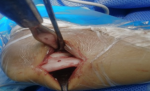
Figure 1: exposition of radioulnar synostosis
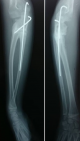
Figure 2: fixation of derotation by 2 K-wire
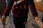
Figure 3: post-operative result of bilateral synostosis
- Ramachandran M, Lau K, Jones DHA. Rotational osteotomies for congenital radioulnar synostosis. J Bone Joint Surg Br. 2005;87(10):1406-1410. PubMed | Google Scholar
- Wilkie DPD. Congenital radio-ulnar synostosis. Br J Surg. 1914. Google Scholar
- Pasupathy B, Tholgappiyan T, Sureshbabu M. Congenital radio ulnar synostosis, analysis of functional outcome using double rotation osteotomy and osteotomy at synostosis site. International Journal of Orthopaedics Sciences. 2018;4(2k):755-759. Google Scholar
- Cleary JE, Omer GE. Congenital proximal radio-ulnar synostosis. Natural history and functional assessment. J Bone Joint Surg Am. 1985;67(4):539-545. PubMed | Google Scholar
- Mital MA. Congenital radioulnar synostosis and congenital dislocation of the radial head. Orthop Clin North Am. 1976;7(2):375-383. PubMed | Google Scholar
- Semaya Ahmed ElSayed. Derotation osteotomy for congenital radioulnar synostosis. Egypt Orthop J. 2014;49(2):92-95. Google Scholar
- Hwang JH, Kim HW, Lee DH, Chung JH, Park H. One-stage rotational osteotomy for congenital radioulnar synostosis. J Hand Surg Eur Vol. 2015;40(8):855-861. PubMed | Google Scholar
- Murase T, Tada K, Yoshida T, Moritomo H. Derotational osteotomy at the shafts of the radius and ulna for congenital radioulnar synostosis. J Hand Surg Am. 2003;28(1):133-137. PubMed | Google Scholar
- Satake H, Kanauchi Y, Kashiwa H, Ishigaki D, Takahara M, Takagi M. Long-term results after simple rotational osteotomy of the radius shaft for congenital radioulnar synostosis. J Shoulder Elbow Surg. 2018;27(8):1373-1379. PubMed | Google Scholar
- Funakoshi T, Kato H, Minami A, Suenaga N, Iwasaki N. The use of pedicled posterior interosseous fat graft for mobilization of congenital radioulnar synostosis: a case report. J Shoulder Elbow Surg. 2004;13(2):230-234. PubMed | Google Scholar
- Hansen OH, Andersen NO. Congenital radio-ulnar synostosis. Report of 37 cases. Acta Orthop Scand. 1970;41(3):225-230. PubMed | Google Scholar
- Sachar K, Akelman E, Ehrlich MG. Radioulnar synostosis. Hand Clin. 1994;10(3):399-404. PubMed
- Shingade VU, Shingade RV, Ughade SN. Results of single-staged rotational osteotomy in a child with congenital proximal radioulnar synostosis: subjective and objective evaluation. J Pediatr Orthop. 2014;34(1):63-69. PubMed | Google Scholar
- Simcock X, Shah AS, Waters PM, Bae DS. Safety and Efficacy of Derotational Osteotomy for Congenital Radioulnar Synostosis. J Pediatr Orthop. 2015;35(8):838-843. PubMed | Google Scholar
- Hung NN. Derotational osteotomy of the proximal radius and the distal ulna for congenital radioulnar synostosis. J Child Orthop. 2008;2(6):481-489. PubMed | Google Scholar
- Kanaya F, Ibaraki K. Mobilization of a congenital proximal radioulnar synostosis with use of a free vascularized fascio-fat graft. J Bone Joint Surg Am. 1998;80(8):1186-1192. PubMed | Google Scholar
- Oka K, Doi K, Suzuki K, Murase T, Goto A, Yoshikawa H et al. In vivo three-dimensional motion analysis of the forearm with radioulnar synostosis treated by the kanaya procedure. Journal of Orthopaedic Research. 2006;24(5):1028-1035. PubMed | Google Scholar
- Castello JR, Garro L, Campo M. Congenital radioulnar synostosis. Surgical correction by derotational osteotomy. Ann Chir Main Memb Super. 1996;15(1):11-17. PubMed | Google Scholar
- Fujimoto M, Kato H, Minami A. Rotational osteotomy at the diaphysis of the radius in the treatment of congenital radioulnar synostosis. Journal of Pediatric Orthopaedics. 2005;25(5):676-679. PubMed | Google Scholar
- El-Adl W. Two-stage double-level rotational osteotomy in the treatment of congenital radioulnar synostosis. Acta Orthop Belg. 2007;73(6):704-709. PubMed | Google Scholar
- Simmons BP, Southmayd WW, Riseborough EJ. Congenital radioulnar synostosis. J Hand Surg Am. 1983;8(6):829-838. PubMed | Google Scholar
- Ogino T, Hikino K. Congenital radio-ulnar synostosis: compensatory rotation around the wrist and rotation osteotomy. J Hand Surg Br. 1987;12(2):173-178. PubMed | Google Scholar
- Khalil I, Vizkelety T. Osteotomy of the synostosis mass for the treatment of congenital radio-ulnar synostosis. Arch Orthop Trauma Surg. 1993;113(1):20-22. PubMed | Google Scholar
- Green WT, Mital MA. Congenital radio-ulnar synostosis: surgical treatment. J Bone Joint Surg Am. 1979;61(5):738-743. PubMed | Google Scholar
- Hankin FM, Smith PA, Kling TF, Louis DS. Ulnar nerve palsy following rotational osteotomy of congenital radioulnar synostosis. J Pediatr Orthop. 1987;7(1):103-106. PubMed | Google Scholar
- Griffet J, Berard J, Michel CR, Caton J. Les synostoses congénitales radio-cubitales supérieures. International Orthopaedics. 1986;10(4):265-269. Google Scholar
- Yammine K, Salon A, Pouliquen JC. Congenital radioulnar synostosis: study of a series of 37 children and adolescents. Annales de Chirurgie de la Main et du Membre Supérieur. 1998;17(4):300-308. PubMed | Google Scholar
Search
This article authors
On Pubmed
On Google Scholar
Citation [Download]
Navigate this article
Similar articles in
Key words
Tables and figures
Article metrics

