
Snare polypectomy in pedunculated colonic polyp
Fouad Nejjari, Aziz Aourarh
Corresponding author: Fouad Nejjari, Gastroenterology Unit, 5th Military Hospital, Guelmim, Morocco 
Received: 21 Nov 2019 - Accepted: 01 Dec 2019 - Published: 09 Dec 2019
Domain: Gastroenterology,Internal medicine,Chronic disease prevention
Keywords: Colonoscopy, snare polypectomy, pedunculated colonic polyp
©Fouad Nejjari et al. PAMJ Clinical Medicine (ISSN: 2707-2797). This is an Open Access article distributed under the terms of the Creative Commons Attribution International 4.0 License (https://creativecommons.org/licenses/by/4.0/), which permits unrestricted use, distribution, and reproduction in any medium, provided the original work is properly cited.
Cite this article: Fouad Nejjari et al. Snare polypectomy in pedunculated colonic polyp. PAMJ Clinical Medicine. 2019;1:48. [doi: 10.11604/pamj-cm.2019.1.48.21055]
Available online at: https://www.clinical-medicine.panafrican-med-journal.com//content/article/1/48/full
Images in medicine 

Snare polypectomy in pedunculated colonic polyp
Snare polypectomy in pedunculated colonic polyp
Fouad Nejjari1,&, Aziz Aourarh2
1Gastroenterology Unit, 5th Military Hospital, Guelmim , Morocco, 2Gastroenterology I Unit, Mohamed V Military Teaching Hospital, Mohamed V- Souissi University, Rabat, Morocco
&Corresponding author
Fouad Nejjari, Gastroenterology Unit, 5th Military Hospital, Guelmim, Morocco

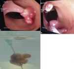
A 55-year-old man with a familial history of colonic neoplasm presented for screening colonoscopy, he had no specific symptomatology, physical examination finding and biological exams were normal. A colonoscopy revealed a pedunculated polyp in the ascending colon with a peduncle of approximately 3cm in length surmounted by a polyp head about 1.5cm in diameter (A), the polyp was identified, the polypectomy snare was passed over the polyp and placed around the middle of the stalk. An electric current was then passed through the snare to cut through the polyp rod (B), thereby providing electrocautery at the same time, then the polyp was recovered using the snare by removing the colonoscope (C). There was no bleeding and no perforation and the patient was discharged within 1 day of endoscopic polypectomy. Anatomopathological examination revealled an adenomatous polyp with high-grade of dysplasia.
Figure 1: (A) endoscopic view of a pedunculated polyp in ascending colon; (B) snare polypectomy with complete resection of the polyp; (C) macroscopic appearance of the polyp after removal from the colon
Search
This article authors
On Pubmed
On Google Scholar
Citation [Download]
Navigate this article
Similar articles in
Key words
Article metrics
PlumX Metrics
Snare polypectomy in pedunculated colonic polyp
