
Fibromatosis colli: about 02 cases
Hassan Doulhousne, Salah Ben Elhend, Nabil Hammoune, Abdelghani El Fikri
Corresponding author: Hassan Doulhousne, Department of Radiology, 5th Military Hospital, Guelmim, Morocco 
Received: 20 Nov 2019 - Accepted: 05 Dec 2019 - Published: 10 Dec 2019
Domain: Public Health Nursing,Pediatrics (general)
Keywords: Fibromatosis colli, sternocleidomastoid, ultrasound
©Hassan Doulhousne et al. PAMJ Clinical Medicine (ISSN: 2707-2797). This is an Open Access article distributed under the terms of the Creative Commons Attribution International 4.0 License (https://creativecommons.org/licenses/by/4.0/), which permits unrestricted use, distribution, and reproduction in any medium, provided the original work is properly cited.
Cite this article: Hassan Doulhousne et al. Fibromatosis colli: about 02 cases. PAMJ Clinical Medicine. 2019;1:51. [doi: 10.11604/pamj-cm.2019.1.51.21042]
Available online at: https://www.clinical-medicine.panafrican-med-journal.com//content/article/1/51/full
Case report 

Fibromatosis colli: about 02 cases
Fibromatosis colli: about 02 cases
Hassan Doulhousne1,&, Salah Ben Elhend2, Nabil Hammoune2, Abdelghani El Fikri1
1Department of Radiology, 5th Military Hospital, Guelmim, Morocco, 2Department of radiology, Military Hospital Avicenna, Marrakech, Morocco
&Corresponding author
Hassan Doulhousne, Department of Radiology, 5th Military Hospital, Guelmim, Morocco
Fibromatosis colli or congenital torticollis is a rare pseudotumor of the sternocleidomastoid muscle of the newborn and infant which the mechanism of occurrence is controversial. The diagnosis is clinical. Ultrasounds make the diagnosis and eliminate other causes of torticollis. The evolution is towards spontaneous regression in a few months or with physiotherapy. We report the collected observations of two infants aged 8 and 12 weeks, respectively presenting a right and left lateral cervical mass. The clinical and ultrasound appearance strongly evoked the diagnosis. The follow-up was ultrasonographic with good clinical evolution and spontaneous regression in a few months.

Fibromatosis colli (FC) (or congenital torticollis) is a rare benign affection of the sternocleidomastoid muscle (SCM) in newborns and infants whose mechanism remains controversial. The imaging (mainly ultrasound) makes a diagnosis and follow-up. The evolution is towards spontaneous regression or under physiotherapy.

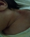
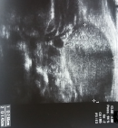
We report two observations of infants aged 8 and 12 weeks who had respectively right and left lateral cervical firm swelling, without inflammatory signs (Figure 1). Ultrasound showed fusiform and homogeneous thickening of the SCM, measuring respectively 2,6cm on the right one and 3,3cm on the left one without other associated abnormalities of neighborhood structures (Figure 2). The follow-up was ultrasonographic with spontaneous regression.
Fibromatosis Colli (FC) is a rare pseudotumor of the SCM whose prevalence is estimated at 0.3 - 2% with male predominance. The right side seems to be the most frequently affected (60-75%) [1]. The mechanism of occurrence remains uncertain. Many theories have been put forward, especially those related to ischemic lesions favored by intrauterine fetal malposition or related to traumas of the muscle during a difficult delivery [2, 3]. The diagnosis of FC is evoked clinically in front of lateral cervical swelling and torticollis. Ultrasound (gold exam) makes the diagnosis and eliminates the other causes of lateral cervical masses by showing the classic imaging of the fusiform thickening of the SCM. It ensures also the follow up during the evolution. The inaccessibility, the cost or the radiating nature of the other means of imaging (CT, MRI) makes that their use is not the routine [3, 4]. The evolution is towards spontaneous regression in 4 to 6 months, facilitated or accelerated by physiotherapy [3-5].
Fibromatosis Colli is a relatively rare pseudotumor of newborns and infants. Diagnosis is evoked clinically. Ultrasound eliminates other causes of torticollis and makes the follow-up. The evolution is towards spontaneous regression or under physiotherapy in a few months.
The authors declare no competing interest.
All the authors have read and agreed to the final manuscript.
Figure 1: left lateral cervical swelling with no inflammatory sign
Figure 2: ultrasound appearance of homogeneous fusiform thickening of the left SCM muscle
- Garetier M, Breton S, Pennaneach A, Barberot C, Chinellato S, Rousset J. Fibromatosis colli. Presse Médicale. 2012;41(2): 213-214. PubMed | Google Scholar
- Kumar B, Pradhan A. Diagnosis of sternomastoid tumor of infancy by fine-needle aspiration cytology. Diagn Cytopathol. 2011 Jan;39(1):13-7. PubMed | Google Scholar
- Mazamaesso Tchaou, Plaodezina Essobozou Pegbessou, Lantam Sonhaye, Patricia Yekpe Ahouanssou, Abdoulatif Amadou, Beresa Kolou et al. Le fibromatosis colli ou torticolis congénital: son diagnostic et sa prise en charge à propos de deux cas. The Pan African Medical Journal. 2015;22:74. PubMed | Google Scholar
- Smiti S, Kulkarni NM, Singh J. Case report: fibromatosis colli in a neonate. Indian J Radiol Imaging. 2010 February;20(1):45-46. PubMed | Google Scholar
- Cheng JC, Wong MW, Tang SP, Chen TMK, Shum SLF, Wong EMC. Clinical determinants of the outcome of manual stretching in the treatment of congenital muscular torticollis in infants. A prospective study of 821 cases. J Bone Joint Surg Am. 2001 May;83(5):679-87. PubMed | Google Scholar
Search
This article authors
On Pubmed
On Google Scholar
Citation [Download]
Navigate this article
Similar articles in
Key words
Tables and figures
Article metrics
PlumX Metrics
Fibromatosis colli: about 02 cases
