
Sigmoid volvulus in pictures: from the exam table to the operating table
Atef Mejri, Mohamed Firas Ayadi
Corresponding author: Atef Mejri, General Surgery Department, Regional Hospital of Jendouba, Jendouba, Tunisia 
Received: 11 Nov 2019 - Accepted: 15 Nov 2019 - Published: 26 Dec 2019
Domain: General surgery
Keywords: Sigmoid, volvulus, surgery, radiology, exam
©Atef Mejri et al. PAMJ Clinical Medicine (ISSN: 2707-2797). This is an Open Access article distributed under the terms of the Creative Commons Attribution International 4.0 License (https://creativecommons.org/licenses/by/4.0/), which permits unrestricted use, distribution, and reproduction in any medium, provided the original work is properly cited.
Cite this article: Atef Mejri et al. Sigmoid volvulus in pictures: from the exam table to the operating table. PAMJ Clinical Medicine. 2019;1:70. [doi: 10.11604/pamj-cm.2019.1.70.20941]
Available online at: https://www.clinical-medicine.panafrican-med-journal.com//content/article/1/70/full
Images in medicine 

Sigmoid volvulus in pictures: from the exam table to the operating table
Sigmoid volvulus in pictures: from the exam table to the operating table
Atef Mejri1,&, Mohamed Firas Ayadi1
1General Surgery Department, Regional Hospital of Jendouba, Jendouba, Tunisia
&Corresponding author
Atef Mejri, General Surgery Department, Regional Hospital of Jendouba, Jendouba, Tunisia

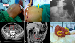
Sigmoid volvulus is a frequent cause of large bowl obstruction. If it is not recognized in time, it can be hazardous with a high risk of mortality. Otherwise, simple exams could help to reach the correct diagnosis in time, which may completely change the prognosis and even avoid surgery in some cases. A 58-year-old man presented to the emergency room with acute abdominal pain associated with distal intestinal obstruction syndrome. The physical examination revealed a remarkable distention of the abdomen and the sigmoid colon whose limits could be discerned under the skin since the patient was thin (A) as well as a depression at the left iliac fossa (B). The plain abdominal radiography showed the « coffee bean » sign (C). A CT-scan was performed showing a « whirl sign » (D,E). The patient underwent emergency surgery. Intraoperatively, the sigmoid colon was markedly dilated and volvulated (F). We performed a resection of the sigmoid colon and primary anastomosis with simple outcomes. Through this case, we can conclude the multiple sings of sigmoid volvulus like the pathognomonic triad of Von Wahl, present in 68.84% of cases. The CT scan permits to confirm the diagnosis by showing the whirl sign: twisting of the mesenteric vessels and search for complications. Regarding the risk of recurrence of 18-90% for the endoscopic decompression, the most considered treatment is sigmoid non-surgical decompression followed by an elective sigmoid resection.
Figure 1: (A) markedly distended sigmoid colon, found especially in the skinny patient; (B) depression at the left iliac fossa (C) plain abdominal radiography: double loop obstruction that shows the « coffee bean »; (D) axial CT scan whirl sign that defines an axial mesenteric volvulus of the sigmoid; (E) multiplanar reconstruction of the CT scan whirl sign; (F) intraoperatively: sigmoid colon: markedly distended and volvulated
Search
This article authors
On Pubmed
On Google Scholar
Citation [Download]
Navigate this article
Similar articles in
Key words
Article metrics

