
Pneumosinus dilatans associated with meningioma
Maria Inês de Sá, Filipa Proença
Corresponding author: Maria Inês de Sá, Neuroradiology, Department of Neuroradiology, Santa Maria Hospital, North Lisboa University Hospital Center, Lisboa, Portugal 
Received: 27 Dec 2022 - Accepted: 14 Jan 2023 - Published: 17 Jan 2023
Domain: Radiology
Keywords: Meningiomas, pneumosinus dilatans, ethmoid sinus
©Maria Inês de Sá et al. PAMJ Clinical Medicine (ISSN: 2707-2797). This is an Open Access article distributed under the terms of the Creative Commons Attribution International 4.0 License (https://creativecommons.org/licenses/by/4.0/), which permits unrestricted use, distribution, and reproduction in any medium, provided the original work is properly cited.
Cite this article: Maria Inês de Sá et al. Pneumosinus dilatans associated with meningioma. PAMJ Clinical Medicine. 2023;11:22. [doi: 10.11604/pamj-cm.2023.11.22.38660]
Available online at: https://www.clinical-medicine.panafrican-med-journal.com//content/article/11/22/full
Images in clinical medicine 

Pneumosinus dilatans associated with meningioma
Pneumosinus dilatans associated with meningioma
![]() Maria Inês de Sá1,&,
Maria Inês de Sá1,&, ![]() Filipa Proença1
Filipa Proença1
&Corresponding author

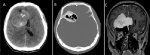
A 54-year-old man presented to the emergency department after having fallen from height. The physical examination was unremarkable. The computed tomographic (CT) brain scan (A, B) showed no signs of trauma, instead, revealed a right frontal solid space-occupying lesion with cystic and calcified areas. Magnetic resonance imaging (MRI) was performed (C) and showed a voluminous dural-based mass, with well-defined borders and a strong and homogeneous contrast-enhancing on T1-WI. The calcified and cystic areas were osteosclerotic reactions and extensive hyperpneumatization of the adjacent ethmoid sinus. Meningioma with associated complex pneumosinus dilatans (PSD), with new sinus cell formation, was the suggested diagnosis. Although it is rare, PSD can occur associated to meningiomas and the theory behind it is obstruction of the sinus ostium causing expansion through a “ball-valve” effect. Effective treatment can avoid pneumocephalus formation. The patient underwent surgical resection, and the histologic and intraoperative observation confirmed the diagnosis.
Figure 1: computed tomographic (CT) brain scan (A, B) revealed a right frontal solid space-occupying lesion with cystic and calcified areas; magnetic resonance imaging (MRI) (C) showed a voluminous dural-based mass, with well-defined borders, a strong and homogeneous contrast-enhancing on T1-WI
Search
This article authors
On Pubmed
On Google Scholar
Citation [Download]
Navigate this article
Similar articles in
Key words
Article metrics
PlumX Metrics
Pneumosinus dilatans associated with meningioma
