
Bladder exstrophy-epispadias complex
Gopidi Sainidhi Reddy, Harshith Gowda
Corresponding author: Harshith Gowda, Department of Radiodiagnosis, Jawaharlal Nehru Medical College, Sawangi (Meghe), Wardha, Maharashtra, India 
Received: 06 Sep 2022 - Accepted: 15 Feb 2023 - Published: 17 Feb 2023
Domain: Pediatrics (general),Pediatric surgery,Urology
Keywords: Bladder, congenital defect, surgery
©Gopidi Sainidhi Reddy et al. PAMJ Clinical Medicine (ISSN: 2707-2797). This is an Open Access article distributed under the terms of the Creative Commons Attribution International 4.0 License (https://creativecommons.org/licenses/by/4.0/), which permits unrestricted use, distribution, and reproduction in any medium, provided the original work is properly cited.
Cite this article: Gopidi Sainidhi Reddy et al. Bladder exstrophy-epispadias complex. PAMJ Clinical Medicine. 2023;11:40. [doi: 10.11604/pamj-cm.2023.11.40.37220]
Available online at: https://www.clinical-medicine.panafrican-med-journal.com//content/article/11/40/full
Images in clinical medicine 

Bladder exstrophy-epispadias complex
Bladder exstrophy-epispadias complex
![]() Gopidi Sainidhi Reddy1,
Gopidi Sainidhi Reddy1, ![]() Harshith Gowda1,&
Harshith Gowda1,&
&Corresponding author

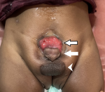
A 2-year-old male, with the bladder exstrophy-epispadias complex since birth, presents to the department of surgical pediatrics for surgical management. Exposed, everted bladder template is clearly obvious immediately below the umbilicus; left and right corpora cavernosa are visible beneath and alongside the urethral plate; the scrotum is caudally displaced. A spectrum of congenital defects' epispadias, typical bladder exstrophy, and cloacal exstrophy, make up the exstrophy-epispadias complex (EEC). Nelson et al. calculated the total incidence of EEC at 2.15 per 100,000 live births, with an equal male-to-female ratio. Normal invagination of the urogenital system begins at the end of the third week of pregnancy in the intermediate layer of mesoderm, while the lateral plate mesoderm contributes to the formation of the primitive gut tube. EEC develops because of disruption in this interaction, which may be caused by an overgrowth of the cloacal membrane that prevents mesenchymal tissue from migrating medially. The severity of the resulting condition depends on the point at which the interaction between the mesodermal layers is disturbed. Depending on the form and degree of the defect, different surgical procedures are required to rectify it; most neonates, however, require bladder and abdominal wall closure, epispadias repair, ureteral reimplantation, and bladder neck repair.
Figure 1: bladder exstrophy (black and white arrow), epispadias (white arrow) and inferiorly displaced scrotum (white arrow head)
Search
This article authors
On Pubmed
On Google Scholar
Citation [Download]
Navigate this article
Similar articles in
Key words
Tables and figures
Article metrics
PlumX Metrics
Bladder exstrophy-epispadias complex
