
Retinal veno-venous anastomosis in chronic branch retinal vein occlusion: when natural healing takes place
Rida El Hadiri, Rim El Hachimi
Corresponding author: Rida El Hadiri, Université Mohammed V de Rabat, Centre Hospitalier Universitaire Ibn Sina, Hôpital des Spécialités, Ophtalmologie A, Rabat, Morocco 
Received: 06 Dec 2021 - Accepted: 02 Mar 2023 - Published: 03 Mar 2023
Domain: Ophthalmology
Keywords: Retinal veno-venous anastomosis, collaterals of retinal vessels, branch retinal vein occlusion
©Rida El Hadiri et al. PAMJ Clinical Medicine (ISSN: 2707-2797). This is an Open Access article distributed under the terms of the Creative Commons Attribution International 4.0 License (https://creativecommons.org/licenses/by/4.0/), which permits unrestricted use, distribution, and reproduction in any medium, provided the original work is properly cited.
Cite this article: Rida El Hadiri et al. Retinal veno-venous anastomosis in chronic branch retinal vein occlusion: when natural healing takes place. PAMJ Clinical Medicine. 2023;11:49. [doi: 10.11604/pamj-cm.2023.11.49.32719]
Available online at: https://www.clinical-medicine.panafrican-med-journal.com//content/article/11/49/full
Images in clinical medicine 

Retinal veno-venous anastomosis in chronic branch retinal vein occlusion: when natural healing takes place
Retinal veno-venous anastomosis in chronic branch retinal vein occlusion: when natural healing takes place
![]() Rida El Hadiri1,&,
Rida El Hadiri1,&, ![]() Rim El Hachimi1
Rim El Hachimi1
&Corresponding author

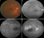
It was a 63-year-old woman with a positive history of systemic hypertension consulting for a three years history of blurred vision Oculus Dexter (OD). She noticed a slowly progressive spontaneous improvement and denied any previous ophthalmology consultation. Best-corrected visual acuity was 6/18 OD with unremarkable adnexal and anterior segment examination. Fundus examination showed supero-temporal branch retinal vein occlusion with arterio-venous nicking, macular exudates, and numerous retinal veno-venous anastomoses bridging the horizontal raphe temporally. The fellow eye was normal. We ordered a fluorescein angiography with color and monochromatic photography that highlighted the previous findings and showed macular diffusion in early and late phases (A,B,C,D). We retained the diagnosis of retinal venovenous anastomosis in chronic branch retinal vein occlusion with chronic macular edema. The patient preferred observation of her condition and refused further investigations or interventions. After two years of follow-up, her visual acuity was stable. We insisted on medical control of her systemic hypertension.
Figure 1: fundus photography showing supero-temporal branch retinal vein occlusion with: A) macular exudates and numerous retinal veno-venous anastomoses; B) highlighted by red-free monochromatic photography; C) fundus fluorescein angiography showed macular diffusion in early; D) late phases
Search
This article authors
On Pubmed
On Google Scholar
Citation [Download]
Navigate this article
Similar articles in
Key words
Article metrics

