
Skin nodules revealing leukemia: observation of a 3-month-old infant
Yousra El Boussaadni, Abdallah Oulmaati
Corresponding author: Yousra El Boussaadni, Pediatrics Service, Faculty of Medicine and Pharmacy, University of Abdel Malek Essaadi, Tangier, Morocco 
Received: 28 Jun 2022 - Accepted: 03 Jan 2023 - Published: 28 Mar 2023
Domain: Pediatric hematology,Pediatric oncology,Pediatrics (general)
Keywords: Skin nodules, leukemia, child
©Yousra El Boussaadni et al. PAMJ Clinical Medicine (ISSN: 2707-2797). This is an Open Access article distributed under the terms of the Creative Commons Attribution International 4.0 License (https://creativecommons.org/licenses/by/4.0/), which permits unrestricted use, distribution, and reproduction in any medium, provided the original work is properly cited.
Cite this article: Yousra El Boussaadni et al. Skin nodules revealing leukemia: observation of a 3-month-old infant. PAMJ Clinical Medicine. 2023;11:54. [doi: 10.11604/pamj-cm.2023.11.54.36110]
Available online at: https://www.clinical-medicine.panafrican-med-journal.com//content/article/11/54/full
Images in clinical medicine 

Skin nodules revealing leukemia: observation of a 3-month-old infant
Skin nodules revealing leukemia: observation of a 3-month-old infant
![]() Yousra El Boussaadni1,&, Abdallah Oulmaati1
Yousra El Boussaadni1,&, Abdallah Oulmaati1
&Corresponding author

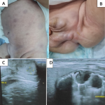
A 3-month-old boy, with no notable pathological history, was brought to the emergency with a history of incessant cry and stool with blood. The diagnosis of intussusception is made after ultrasound. After surgical reduction, exploration found a mesenteric node with a hyperplastic character on the anatomopathological study. The postoperative time was simple, and the child was seen in consultation 15 days after discharge. The examination notes dermo-hypodermic lumps on the trunk, back and testicles, an abdominal ultrasound is requested returning with gastric thickening, periportal infiltration and testicles suggesting lymphoma. The biopsy of the belly lump showed infiltrated hypodermis, microscopically, the tumor cells were round. The cytoplasm was scarce. The nuclei were round, oval or focally irregular, and the mitosis was visible. The neoplasms were positive for Pax5, CD45, CD79 by immunohistochemical staining, which suggests leukemia or lymphoma. The blood count is normal, bone aspiration confirmed the final diagnosis of ALL-B with chloromas. The child receives chemotherapy according to the care protocol of the unfavorable risk group, with a follow-up of 12 months.
Figure 1: A) hard skin lumps all over the body; B) nodules on both testicles measuring 2 cm; C) hypoechoic testicular nodules measuring 14x9x12.5mm on the right and 4.5x5x7.5mm on the left on ultrasound; D) hypoechoic nodular gastric thickening of 12x9X10mm on ultrasound
Search
This article authors
On Pubmed
On Google Scholar
Citation [Download]
Navigate this article
Similar articles in
Key words
Article metrics

