Molecular characterization of genital human papillomavirus among women in Yaoundé, Cameroon
Cynthia Malung Mbimenyuy, Jerome Fru Cho, Akongnwi Emmanuel Mugyia, George Mondinde Ikomey, Denis Manga Tebit, Damian Anong Nota
Corresponding author: Cynthia Malung Mbimenyuy, Department of Microbiology and Parasitology, Faculty of Science, P. O. Box 63, University of Buea, Buea, South West Region, Cameroon 
Received: 12 Oct 2023 - Accepted: 09 Jul 2024 - Published: 05 Aug 2024
Domain: Microbiology, Virology
Keywords: Human papillomavirus (HPV), genotypes, neoplasia, phylogenetic tree, cervical intra-epithelial neoplasia (CIN), oncogenic
©Cynthia Malung Mbimenyuy et al. PAMJ Clinical Medicine (ISSN: 2707-2797). This is an Open Access article distributed under the terms of the Creative Commons Attribution International 4.0 License (https://creativecommons.org/licenses/by/4.0/), which permits unrestricted use, distribution, and reproduction in any medium, provided the original work is properly cited.
Cite this article: Cynthia Malung Mbimenyuy et al. Molecular characterization of genital human papillomavirus among women in Yaoundé, Cameroon. PAMJ Clinical Medicine. 2024;15:32. [doi: 10.11604/pamj-cm.2024.15.32.41951]
Available online at: https://www.clinical-medicine.panafrican-med-journal.com//content/article/15/32/full
Research 
Molecular characterization of genital human papillomavirus among women in Yaoundé, Cameroon
Molecular characterization of genital human papillomavirus among women in Yaoundé, Cameroon
Cynthia Malung Mbimenyuy1,&, Jerome Fru Cho1, ![]() Akongnwi Emmanuel Mugyia2, George Mondinde Ikomey3, Denis Manga Tebit4,5, Damian Anong Nota6
Akongnwi Emmanuel Mugyia2, George Mondinde Ikomey3, Denis Manga Tebit4,5, Damian Anong Nota6
&Corresponding author
Introduction: cervical infection with human papillomavirus (HPV) is mostly associated with cervical neoplasia and these viruses cause a broad scope of diseases from benign to invasive neoplasia. Human papillomavirus types 16, 18, and 45 account for more than 90% of HPV-related cervical intraepithelial lesions in women. This study aimed to evaluate the predominant oncogenic genotypes circulating in Cameroonian women and more specifically to identify the molecular characterization of HPV types 16, 18, and 45.
Methods: a cross-sectional and analytical study was carried out from November 2019 to January 2021 at the Yaoundé General Hospital and the Yaoundé Gyneco-obstetric and Pediatric Hospital. Genomic DNA was extracted and identified from cervical smears by conventional polymerase chain reaction method (PCR). The genotyping was performed using the Sanger sequencing method with the consensus primers. The descriptive variables were statistically analyzed by Chi-square non-parametric test; p-value ≤ 0.05 was noted as statistically associated.
Results: the frequently identified HPV genotypes were HPV-16 (40.6%), HPV-45 (15.6%), and HPV-18 (7.8%) respectively. For HPV16 and 45, the phylogenetic tree corresponding to the L1 ORF region showed that these isolates constitute a new clade as they did not cluster around any of the isolates or lineages from the Gene Bank. Just one HPV18 isolate clustered closely or has a kinship with African sub-lineage (B1) and sub-lineage from other African countries.
Conclusion: results from this study confirmed the burden and involvement of HPV 16, 45, and 18 in cervical intraepithelial lesions and subsequently cervical cancer in Cameroonian women. It further increases doubt on the efficacy and potential benefit of the available HPV vaccines in Cameroon.
Cervical infections caused by oncogenic human papillomavirus (HPV) are mostly sexually transmitted amongst sexually active individuals. HPV has demonstrated differential oncogenic potential and has been associated with cervical cancer (CC) that manifests as two major histologic types; squamous cell carcinoma (SCC) and adenocarcinoma (AC) [1,2]. Human papillomavirus are non-enveloped, circular double-stranded DNA and epitheliotropic viruses with approximately 8,000 base pairs [3]. The genome encodes for two late viral capsid proteins (L1-major and L2-minor) with their main functions in viral structure formation and six early proteins (E1, E2, E4, E5, E6, and E7) for viral replication [4]. Approximately, more than 180 HPV types have been identified and studied based on these late (L) and early (E) sequences with established proofs of sequence variations in some genes of the different genotypes [5] but the L1 genes are mostly used because of their genetic stability.
Oncogenic HPV has been linked to almost all cervical cancer cases globally, ranking it the 4th most common cancer among women. In Cameroon, about 2,770 new cases are diagnosed annually, ranking it the second cause of cancer morbidity and mortality in women nationwide in this setting [6]. Human papillomavirus sequence variability and divergence in sub-Saharan Africa are very high [7], and genotypic variations in the circulating HPV types have been noted in women in different parts of Cameroon. Consequently, this has raised concerns about the effectiveness of the presently available HPV vaccines in the country [6].
Studies have proven that HPV types 16, 18, and 45 are causally linked to more than 80% of cervical cancer globally [8], but HPV 16 dominates in almost all squamous cell carcinoma while HPV18 is mostly identified in adenocarcinoma [9]. It has been noted that the rate of cervical neoplasia attributed to oncogenic or high risk (HR) HPV is elevated in Africa (31.5/100,000 women/year), and more specifically in sub-Saharan Africa (75.3/100,000 women/year). This observed increased rate of HR-HPV in developing countries has been associated with a lack of healthcare accessibility, limited screening programs, and a low rate of HPV immunization [10]. The objectives of this study were to determine the circulating oncogenic HPV genotypes and study their molecular characterization, thereby providing information on their sequence diversity or relatedness to develop effective HPV immunization strategies and control measures.
Study design and setting: this was a cross-sectional and analytical study carried out from November 2019 to January 2021 at the Yaoundé General Hospital and the Yaoundé Gyneco-obstetric and Pediatric Hospital. Recruitment of subjects and data collection was done in the Department of Gynecology and Oncology unit, of the above two mentioned hospitals.
Participants: the recruited participants were women aged, 19 years and older, sexually active, experiencing signs and symptoms of cervical precancerous lesions, and also HIV-positive women coming for routine gynecological consultation. Women were exempted from this study, if they experienced contra-indication for Pap smear examination, and were unwilling to take part.
Variables: in this cross-sectional study, the exposure of the HIV-positive and HIV-negative population to HPV infection was investigated. The dependent variables were incident HPV-related cervical lesions/infection, and these dependent variables were the study's primary outcomes. The following independent variables were extracted from the patient's medical file, questionnaires, and laboratory test results.
Data: the sources of data were the recruited participants, laboratory results, and medical files.
Study size: a minimum sample size of 226 women was calculated using the statistical (Lorentz) formula for proportion:

Where N is the minimum number of participants, and p is the pathological prevalence of HPV=39.0% (p=0.39) in Cameroonian women in 2016 by Catarino et al. [11]. Zα is the 95% confidence interval (Zα=1.96), and d is the error rate set at 5% (d=0.05).
Data collection procedures: the ethical research and review committee of the hospital approved the study protocol. The study and informed consent were explained to patients either in English, French, or a local Lingua franca. After signing the consent form, a questionnaire was given to each study participant for data collection. Patients were tested if they showed no proof of their HIV status.
Cervical cytology sample collection and deoxyribonucleic acid extraction: an endocervical brush (Cytyc, Montrouge, France) was used for cervical sample collection and then stored in a commercial aqueous buffered specimen collection and transport media (STM) (Roche Diagnostic Systems, Meylan, France). All the specimens were stored at -20°C, pending DNA extraction and amplification. Cervical cytology samples were processed at the Centre for the Study and Control of Communicable Diseases (CSCCD) of the Faculty of Medicine and Biomedical Sciences, University of Yaoundé 1. Deoxyribonucleic acid of cervicovaginal samples was extracted using Qiagen DNeasy Kit (Hamburg, Germany; Cat no: 66404) following the manufacturer´s instructions. The extracted DNA was quantified using a Nanodrop 200°C spectrophotometer (Thermo Scientific, Loughborough, UK). The extracted DNA was aliquoted in 50μl aliquots in sterile Eppendorf tubes and stored at -20°C.
Primers and sequences: all primers used in this study shown below, were synthesized in South Africa by Integrated DNA Technologies, (IDT) Inc, and have been described in previous published studies [12].
Polymerase chain reaction amplification: the puReTaq™ Ready-To-Go™ polymerase chain reaction (PCR) beads from Cytiva are designed with high-quality buffer, nucleotides (dNTPs), and thermal-stable recombinant puReTaq DNA polymerase, except primer, DNA template, and water. Polymerase chain reaction amplifications were performed in a 25μL volume containing 21μl of PCR grade water, 1μl forward primer,1μl reverse primer and 2μl DNA template with the thermal cycler, BIO-RAD T100.
Deoxyribonucleic acid extraction control: all the samples were first analyzed using the primer set; CYP2C8 and this primer set amplifies the human gene, CYP2C8 (Cytochrome P450 Family 2 Subfamily C Member 8). These housekeeping genes are constantly expressed in all conditions [13]. We decided to amplify a relatively short portion of the human CYP2C8 gene as a DNA extraction control to obtain a 107bp amplicon with the forward and reverse primers (CYP2C8-F and CYP2C8-R). The thermal cycling condition consisted of 40 cycles of denaturation at 95°C for 30 seconds, annealing at 55°C for 60 seconds, elongation at 72°C for 2 minutes, and a final extension step at 72°C for 5 minutes. Samples that were negative for CYP2C8 genes were eliminated. After succeeding with the control run above, we now proceeded with the HPV-PCR which amplified the L1 gene. The primers listed in Table 1, were used in two rounds of nested PCR, all targeting the L1 conserved region of the HPV genome in the listed order below. Amplification results with the CYP2C8 primers are shown in Figure 1.
First round: SB01 (forward) and SB02 (reverse) or outer polymerase chain reaction analysis: human papillomavirus DNA samples were analyzed in the first round of reaction using SB01/02 PCR primers adapted from Coser et al. 2011 [14]. Thermal cycling conditions for outer PCR were as follows: pre-denaturation; 95°C for 5 minutes, denaturation; 95°C for 30 seconds, annealing; 42°C for 45 seconds, extension; 72°C for 1 minute, and final extension 72°C for 10 minutes. The PCR product was approximately 495bp for the majority of HPV genotypes. Negative controls and molecular weight markers were included with all PCR series and were analyzed in parallel with the clinical specimens (Figure 2).
Second round: MY09 (forward) and MY11 (reverse) or nested polymerase chain reaction analysis: the second round of PCR amplification with the same samples analyzed in the first round of reaction was carried out with consensus primers MY09/MYII [15]. Thermal cycling conditions were adopted from Cheng et al. and were as follows: pre-denaturation; 95°C for 5 minutes, denaturation; 95°C for 15 seconds, annealing; 50°C for 1 minute, extension; 72°C for 1 minute, and final extension 72°C for 10 minutes. Negative samples were added as controls in all the experiments for quality control. Amplicons underwent electrophoresis in agarose gel and positive and negative results were evaluated based on the presence of fragments of the expected size (450bp) (Figure 3).
Gel electrophoresis: all PCR amplified products were resolved in 2% agarose gel (Sigma Aldrich) dissolved in a 1X Tris-acetic EDTA (TAE) buffer and the gel electrophoresis was run at 100 volts for 60 minutes. The amplified PCR products were visualized under ultra violet (UV) light using a trans-illuminator. The samples that tested HPV-DNA positive in the nested PCR were analyzed for specific genotypes by Sanger sequencing.
Sequence analysis: the amplified DNA samples were sent for sequencing to Inqaba Biotec, West Africa Ltd Ibadan Nigeria using prepaid barcodes. DNA sequencing was performed by the Sanger sequencing technique. Each PCR tube sent for sequencing contained 15μL of the DNA template and 100μl of 25μM concentration of the consensus MY09 and MY11 primers. Finally, sequences obtained were compared with virus sequences available at the National Center for Biotechnology Information (NCBI), using the online standard Nucleotide BLAST software.
Phylogenetic analysis and tree construction: molecular Evolutionary Genetics Analysis (MEGA) software version 11.0 was used to identify the evolutionary relationships among the analyzed sequences based on L1 nucleotide sequences and multiple alignments were performed by using the CLUSTALW program [16]. The Neighbor-Joining algorithm and the Kimura 2-parameter model trees [17], with 1000 bootstrapped replicates, were built and Phylogenetic analyses were performed with LCR sequences of HPV16, HPV18, and HPV45. The obtained isolates were aligned against reference sequences with the following NCBI GenBank database with accession number found in Table 2.
Statistical analysis: all data were analyzed, using SPSS Version 16 (SPSS, Chicago, IL, USA). The descriptive variables were statistically analyzed by Chi-square non-parametric test; P < 0.05 was considered statistically significant. The statistical comparison was performed between high risk (HR), low risk (LR), and unclassified risk (UR).
Data handling: data of laboratory results were double entered in a Microsoft Excel spreadsheet, and any discrepancies were reviewed and corrected using source documents like medical files and laboratory reports.
Ethical consideration and approval: the study was approved by the Research Ethics Committee of the University of Buea. Reference No. 2017/0491/UB/FS/HOD/MBP. Local ethical clearance was also obtained from the Institutional Ethics Committee for Research of Human Health (CIERSH) at the Yaoundé Gynaeco-Obstetric and Pediatric Hospital, Authorization No. 675/CIERSH/DM/2018 and Yaoundé General Hospital, Authorization No. 3616/017/HGY/DG.
Socio-demographic characteristics: a total of 62 women living with HIV and 164 HIV-negative women took part in the study and were diagnosed with cervical lesions or abnormalities. The age range of the participants were between 20 to 79 years, with a median age of 44 years [SD: 10.651]. Most of the women were of age between 40 to 49 years and the majority 131 (57.9%) of them were married, with only 61 (26.9%) who had not attained at least a secondary level of education.
Human papillomavirus detection and genotyping: human papillomavirus-deoxyribonucleic acid detection was carried out on all the 226 cervical specimens collected, from the women who consulted at the two study sites from which, 77 (34.07%) were positive for HPV-DNA with the consensus primers that targeted the L1 open reading frame (ORF) genes. Of the 62 WLWH, 21 (33.8%) tested positive for HPV-DNA and from the 164 HIV-negative women, 56 (34.14%) tested positive for HPV-DNA. Of 77 HPV-DNA positive samples detected by conventional PCR, 50 were sent for genotyping using the Sanger sequencing. Unfortunately, 44 specimens were validated by the quality control procedure for sequencing. From the results of the 44 sequenced samples, 17 individual HPV types were detected at various frequencies, amongst which, 27/44 (61.4%) samples were infected with single HPV genotypes, 14/44 (31.8%) with double/dual infections, and 3/44 (6.8%) with triple infections respectively. Taking into consideration samples with a single HPV infection, HPV-16 was isolated from 20(45.5%) women while we noticed an equal rate of 2 (4.5%) for HPV-18 and HPV-45. With regards to multiple infections, a total of 17 (38.6%) samples were infected by other HPV types, with the majority of 5 (11.5%) samples being positive for HPV16/45 and 2 (4.5%) infected with 18/45/97 (Table 3). Positive samples of HPV16, HPV18, and HPV45 were separated from the rest, for phylogenetic characterization.
The HPV isolates included 11 high-risk types (HPV-16, 18, 31, 33, 35, 45, 51, 52, 58, 68, 82), 5 low-risk types (HPV-13, 54, 62, 72, 81) and one unclassified risk (HPV-96) respectively. High-risk HPV types were more frequently 56 (87.5%) diagnosed among HPV-positive individuals than low-risk types 5 (7.8%). The most commonly detected HR-HPV types were HPV-16, 26 (40.6%), followed by HPV 45 with a frequency of 10 (15.6%), and lastly, HPV 18 with 5 (7.8%). Twenty-six single infections with only the HR genotypes were most frequently diagnosed and this rate was closely followed by 12, HR-HR co-infections (Table 3).
Other analysis
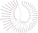
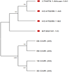
Molecular characterization of the oncogenic human papillomavirus types 16, 18, and 45: variant distribution was determined through L1 ORF sequences. HPV16 isolates were examined for nucleotide variation within a 1707bp-nucleotide (nt) (5430 to 7136). In the phylogenetic tree, nitrogenous bases isolated for all the identified HPV types have bootstrapped more than 90%. This was used to evaluate the feasibility of phylogenetic trees. After examining the sequencing results, of the 44 HPV-positive DNA samples, we detected a high (75%) degree of genetic variability and sequence polymorphism/divergence in our sample (HPV 18 and 45) from the reference sample, with the introduction of four new alleles leading to new haplotypes in our samples. Phylogenetic analysis demonstrated that the distribution of all the isolated HPV genotypes, and particularly phylogenetic relatedness was very divergent (Figure 4). The HPV16 and 45, the phylogenetic tree showed that these isolates constitute a new clade as they did not cluster around any of the isolates or lineages from the Gene Bank, suggesting the possibility of a hitherto or previously undescribed HPV16 and 45 variants (Figure 5, Figure 6).
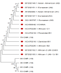
Considering the HPV16 genotype, there was even a greater (92%) percentage of divergence from the reference, with the introduction of five new alleles. The phylogenetic tree corresponding to HPV18 L1 Nitrogen Bases with the Neighbour-Joining method showed that only one HPV18 isolate clustered closely or has a kinship with the African sub-lineage (B1) and other African countries. This suggests that 79CMR could be a recombinant of African variants (Figure 7).
This study was conducted amongst women presenting the signs and symptoms of cervical infection and many of them had been referred from other health facilities following complaints suggestive of either cervical infection or lesions. Therefore, the increased rate of HPV16, HPV 45, and HPV18 diagnosed in these women with precancerous lesions is not alarming, for this group of women is considered a “high risk” group for HPV infection. According to results obtained from these sequenced samples, the frequently detected HPV genotypes that infect Cameroonian women were HPV-16, 26(40.6) followed by HPV-45, 10(15.6%) and HPV-18, 5(7.8%) with a greater proportion of 27(61.4%) of single infections detected and 17(38.6%) multiple infections. Individual infections were more of the HR genotypes, 56(87.5%) than of the LR-genotype, 5(7.8%). A similar result was reported from another study conducted in the same city town by T. Simo et al. 2021, with the following encountered HR-HPV types, HPV-16(54.1%), HPV-18(31.7 %), and HPV-45 (19.5%), both as single infections and multiple infections [18]. Doh et al. 2021 also identified the most prominent HPV types as HPV-16; (24%), HPV-18; (36.4%), and HPV-45; (28%) [19]. In another study, Pirek et al. 2015 noted the following prevalence; HPV-16; (88%), HPV-45; (32%), and HPV-18; (14.8%) in descending order with 54.9% of cases infected with a single HPV type while 45.1% had two or more HPV infections [20].
The distribution of oncogenic HPV types was different in other neighboring countries like Nigeria where Manga et al. 2015 noted that the five most predominant genotypes were 18, 16, 33, 31, and 35, with the prevalence of 44.7%, 13.2%, 7.9%, 5.3% and 5.3% respectively [21]. In a multi-centric study conducted on Togolese women, Kuassi-Kpede et al. 2021 reported that the most common genotypes were HPV-56 (22.7%), followed by HPV-51 (20.3%), HPV-31 (19.5%), HPV-52 (18.8%) and HPV-35 (17.2%) in decreasing order [22]. It is interesting to know that, all of the commonly identified HPV types (16, 18, 31, 33, 35, 45, 52, and 58) cited globally as the causative agent for cervical cancer were also diagnosed in the women recruited in this study [23]. The methodology of this study was based on nested PCR using consensus primer SB01/SB02 and PGMY 09/11 which has demonstrated its effectiveness and sensitivity in detecting a wider range of HPV genotypes and also effective in diagnosing multiple infections [24]. However, in this study, the detection of LR-HPV genotypes was not as effective as with the HR-HPV types. This limitation may be attributed to the fact that PGMY 09/11 primer has a low capacity for detecting low-risk-HPV and could again be related to the fact that, the low-risk types are generally fewer in cervical specimens [25].
This study is the first to date in Cameroon to propose an evolutionary scenario for HPV-16, HPV-18, and HPV-45. The sequencing findings and phylogenetic analysis demonstrated that the distribution of the HPV genotypes, and particularly phylogenetic relatedness was very divergent. From the phylogenetic analysis results obtained from this current study, HPV16 and HPV 45 isolates may constitute a new clade of these two genotypes suggesting a particular virus-host co-evolution in Cameroonian women. Again, all the HPV-18 Isolates diverged from the reference samples except isolate 79CMR which clustered closely or had a kinship with the African sub-lineage (B1) and other African countries. This suggests that 79CMR could be a recombinant of African variants and might reflect the contribution of populations from sub-Saharan African origin in the epidemiology of HPV.
The divergence in the predominance of genotypic and phenotypic variants within HPV types shows that certain types of HPV are clustered according to the ethnic people and regions where they are isolated [26]. This above-mentioned statement can be attributed to the fact that the distribution of HPV types is based on the proximity of the geographical location, ethnicity, and lifestyle of the individual [27]. These consideration makes it possible to doubt or question the effectiveness of the HPV immunization program put in place by the Cameroonian government which is based mostly on Quadrivalent or Gardasil that protect against only 2 of the oncogenic genotypes 16 and 18. While type-specific protection is expected, the perspectives of the administration of prophylactic vaccines highlight the need to reinforce the knowledge of the type-specific prevalence of high-risk HPVs in Cameroon.
Study limitations: a possible limitation of the study was the fact that, the conventional PCR assay used in the study for HPV-DNA detection might have a limited sensitivity level, assuming that a certain proportion of HPV-DNA might have not been detected. The number of samples sent for sequencing was limited, not giving a good representation of the burden of the disease in the country.
It was noticed that three most frequent genotypes diagnosed in HPV-positive subjects were mostly the HR types according to their oncogenic and molecular characterization. This high rate of oncogenic types is an important public health concern. Again, considering the results obtained from this present study, the uniformity or homogeneity in the HPV isolates identified makes it difficult to perform a phylogenetic comparison. Further studies will be necessary to determine whether the HPV16, HPV18 and HPV45 variants identified in this study have a particular oncogenic potential and also to analyze the relationship between variants and their possible impact of the variability observed in ORF L1 on the HPV vaccine efficacy.
What is known about this topic
- Human papillomavirus-16, 18, and 45 are associated with about more than 90% of cervical cancer cases globally, and are the second most frequent gynecological/genital HPV-related cancers in women;
- In Cameroon, about 2,770 new cases are diagnosed annually, ranking it the second cause of female cancer and the second leading cause of cancer deaths;
- Human papillomavirus sequence variability and divergence in sub-Saharan Africa are very high.
What this study adds
- This study identified the three most frequent oncogenic HPV genotypes in Cameroonian women (genotype 16, 45, and 18);
- The sequencing findings and phylogenetic analysis demonstrated that the distribution of the HPV genotypes was divergent.
The authors declare no competing interests.
Cynthia Malung Mbimenyuy conceptualized the study, performed the analysis, interpreted the data, and wrote the manuscript. Denis Manga Tebit drafted, analyzed data and reviewed the manuscript. Jerome Fru Cho performed the experiment and analyzed data. Damian Anong Nota reviewed the manuscript and analyzed the data. Akongnwi Emmanuel Mugyia analyzed the data. All the authors have read and approved the final version of the manuscript.
Table 1: primer sequences, amplicon sizes, and targeted regions
Table 2: human papillomavirus lineages included in the phylogenetic classification of this study
Table 3: types of infection and their related risk types among individuals tested
Figure 1: amplification of HPV DNA template using the control CYP2C8 primer pair; MWM: molecular marker/ladder; (1000bp), NC: negative control: agarose gel (2%) showing PCR amplified DNA fragments of different sizes with CYP2C8 (107bp)
Figure 2: amplification of HPV DNA template using outer PCR (SB01/SB02); MWM: molecular marker (1000bp); NC: negative control; numbers refer to samples; both nested PCR carried out using 3 ml template DNA and PCR amplicons were run on 2.5% agarose gel stained with ethidium bromide
Figure 3: amplification of HPV DNA template using nested PCR (MY09/11); M: molecular marker (1000bp); NC: negative control; numbers refer to samples; both nested PCR carried out using 3 ml template DNA and PCR amplicons were run on 2.5% agarose gel stained with ethidium bromide
Figure 4: phylogenetic tree constructed by the neighbor-joining method based on the genotype sequences of 44 sequenced Cameroonian HPV isolates
Figure 5: phylogenetic trees of HPV16 based on L1 nucleotide constructed using the neighbor-joining method
Figure 6: phylogenetic trees of HPV 45 based on L1 nucleotide constructed using neighbor-joining method
Figure 7: phylogenetic trees of HPV18 based on L1 nucleotide constructed using neighbor-joining method
- Georgieva S, Iordanov V, Sergieva S. Nature of cervical cancer and other HPV-associated cancers. J BUON. 2009 Jul-Sep;14(3):391-8. PubMed | Google Scholar
- Bernard H, Burk R, Chen Z, van Doorslaer K, Hausen H, de Villiers E. Classification of papillomaviruses (PVs) based on 189 PV types and proposal of taxonomic amendments. Virology. 2010;401(1):70-79. PubMed | Google Scholar
- Woodman C, Collins S, Young L. The natural history of cervical HPV infection: unresolved issues. Nat Rev Cancer. 2007 Jan;7(1):11-22. PubMed | Google Scholar
- Fernandes J, Carvalho M, de Fernandes T, Araújo J, Azevedo P, Azevedo J et al. Prevalence of human papillomavirus type 58 in women with or without cervical lesions in northeast Brazil. Ann Med Health Sci Res. 2013 Oct;3(4):504-10. PubMed | Google Scholar
- Chen Z, Schiffman M, Herrero R, DeSalle R, Anastos K, Segondy M et al. Evolution and Taxonomic Classification of Alphapapillomavirus 7 Complete Genomes: HPV18, HPV39, HPV45, HPV59, HPV68 and HPV70. PLoS One. 2013 Aug 16;8(8):e72565. PubMed | Google Scholar
- Bruni L, Albero G, Serrano B, Mena M, Collado JJ, Gómez D et al. ICO/IARC Information Centre on HPV and Cancer (HPV Information Centre). Human Papillomavirus and Related Diseases in Cameroon. Summary Report 22 October 2021.
- Muñoz N, Bosch FX, de Sanjosé S, Herrero R, Castellsagué X, Shah KV et al. Epidemiologic classification of human papillomavirus types associated with cervical cancer. N Engl J Med. 2003 Feb 6;348(6):518-27. PubMed | Google Scholar
- Bruni L, Diaz M, Castellsagué M, Ferrer E, Bosch FX, de Sanjosé S. Cervical human papillomavirus prevalence in 5 continents: Meta-analysis of 1 million women with normal cytological findings. J Infect Dis. 2010 Dec 15;202(12):1789-99. PubMed | Google Scholar
- Tjalma W, Van Waes T, Van den Eeden L, Bogers J. Role of human papillomavirus in the carcinogenesis of squamous cell carcinoma and adenocarcinoma of the cervix. Best Pract Res Clin Obstet Gynaecol. 2005 Aug; 19(4):469-83. PubMed | Google Scholar
- Kombe A, Li B, Zahid A, Mengist HM, Bounda G-A, Zhou Y et al. Epidemiology and Burden of Human Papillomavirus and Related Diseases, Molecular Pathogenesis, and Vaccine Evaluation. Front Public Health. 2021;8:552028. PubMed | Google Scholar
- Catarino Rosa, Vassilakos Pierre, Tebe Pierre-Marie, Schäfer Sonja, Bongoe Adamo, Petignat Patrick. Risk factors associated with human papillomavirus prevalence and cervical neoplasia among Cameroonian women. Cancer Epidemiol. 2016 Feb:40:60-6. PubMed | Google Scholar
- Tawe L, Grover S, Narasimhamurthy M, Moyo S, Gaseitsiwe S, Kasvosve I et al. Molecular detection of human papillomavirus (HPV) in highly fragmented DNA from cervical cancer biopsies using double-nested PCR. MethodsX. 2018 May 31:5:569-578. PubMed | Google Scholar
- Zhu B, Pennack J, McQuilton P, Forero MG, Mizuguchi K, Sutcliffe B et al. Drosophila neurotrophins reveal a common mechanism for nervous system formation. PLoS Biol. 2008;6(11):e284. PubMed | Google Scholar
- Coser J, Boeira Tda R, Fonseca AS, Ikuta N, Lunge VR. Human papillomavirus detection and typing using a nested-PCR-RFLP assay. Braz J Infect Dis. 2011 Sep-Oct;15(5):467-72. PubMed | Google Scholar
- Bah C, Anyanwu M, Wright E, Kimmitt P. Human papillomavirus genotype distribution and risk factor analysis in reproductive age women in urban Gambia. J Med Microbiol. 2018 Nov;67(11):1645-1654. PubMed | Google Scholar
- Tamura K, Stecher G, Kumar S. MEGA 11: Molecular Evolutionary Genetics Analysis Version 11. Mol Biol Evol. 2021 Jun 25;38(7):3022-3027. PubMed | Google Scholar
- Tamura K, Nei M. Estimation of the number of nucleotide substitutions in the control region of mitochondrial DNA in Humans and chimpanzees. Mol Biol Evol. 1993 May;10(3):512-26. PubMed | Google Scholar
- Tagne Simo R, Kiafon F, Nangue C, Goura A, Ebune J, Usani M et al. Influence of HIV infection on the distribution of high-risk HPV types among women with cervical precancerous lesions in Yaounde, Cameroon. Int J Infect Dis. 2021 Sep:110:426-432. PubMed | Google Scholar
- Doh G, Mkong E, Ikomey GM, Obasa AE, Mesembe M, Fokunang C et al. Preinvasive cervical lesions and high prevalence of human papilloma virus among pregnant women in Cameroon. Germs. 2021 Mar 15;11(1):78-87. PubMed | Google Scholar
- Pirek D, Petignat P, Vassilakos P, Gourmaud J, Pache JC, Rubbia-Brandt L et al. Human papillomavirus genotype distribution among Cameroonian women with invasive cervical cancer: a retrospective study. Sex Transm Infect. 2015 Sep;91(6):440-4. PubMed | Google Scholar
- Manga MM, Fowotade A, Abdullahi YM, El-Nafaty AU, Adamu DB, Pindiga HU et al. Epidemiological patterns of cervical human papillomavirus infection among women presenting for cervical cancer screening in North-Eastern Nigeria. Infect Agent Cancer. 2015 Oct 2:10:39. PubMed | Google Scholar
- Kuassi-Kpede AP, Dolou E, Zohoncon TM, Traore IMA, Katawa G, Ouedraogo RA et al. Molecular characterization of high-risk human papillomavirus (HR-HPV) in women in Lomé, Togo. BMC Infect Dis. 2021 Mar 19;21(1):278. PubMed | Google Scholar
- Bruni L, Diaz M, Castellsagué M, Ferrer E, Bosch FX, de Sanjosé S. Cervical Human Papillomavirus Prevalence in 5 Continents: Meta-Analysis of 1 Million Women with Normal Cytological Findings. J Infect Dis. 2010 Dec 15;202(12):1789-99. PubMed | Google Scholar
- Cai YP, Yang Y, Zhu BL, Li Y, Xia XY, Zhang RF et al. Comparison of human papillomavirus detection and genotyping with four different prime sets by PCR-sequencing. Biomed Environ Sci. 2013 Jan;26(1):40-7. PubMed | Google Scholar
- Basu P, Chandna P, Bamezai RN, Siddiqi M, Saranath D, Lear A et al. MassARRAY spectrometry is more sensitive than PreTect HPV-Proofer and consensus PCR for type-specific detection of high-risk oncogenic HPV genotypes in cervical cancer. J Clin Microbiol. 2011 Oct; 49(10):3537-44. PubMed | Google Scholar
- Burk RD, Chen Z, Van Doorslaer K. Human papillomaviruses: Genetic basis of carcinogenicity. Public Health Genomics. 2009;12(5-6):281-90. PubMed | Google Scholar
- Baloch Z, Yasmeen N, Li Y, Ma K, Wu X, Yang S et al. Prevalence and risk factors for human papillomavirus infection among Chinese ethnic women in southern of Yunnan, China. Braz J Infect Dis. 2017 May-Jun;21(3):325-332. PubMed | Google Scholar


