
In ankle injury: don’t overlook the peroneal tendons dislocation
Naoufal Elghoul, Mohamed Zaaf
Corresponding author: Naoufal Elghoul, Orthopedic Surgery and Traumatology, Department of Orthopedic Surgery and Traumatology, Military Hospital Mohammed V (HMIMV),Faculty of Medicine and Pharmacy, Mohammed V University of Rabat, Morocco 
Received: 13 Dec 2019 - Accepted: 04 Feb 2020 - Published: 05 Feb 2020
Domain: Emergency medicine,Sport medicine,Orthopedic surgery
Keywords: Peroneal tendon, ankle sprain, dislocation
©Naoufal Elghoul et al. PAMJ Clinical Medicine (ISSN: 2707-2797). This is an Open Access article distributed under the terms of the Creative Commons Attribution International 4.0 License (https://creativecommons.org/licenses/by/4.0/), which permits unrestricted use, distribution, and reproduction in any medium, provided the original work is properly cited.
Cite this article: Naoufal Elghoul et al. In ankle injury: don’t overlook the peroneal tendons dislocation. PAMJ Clinical Medicine. 2020;2:34. [doi: 10.11604/pamj-cm.2020.2.34.21302]
Available online at: https://www.clinical-medicine.panafrican-med-journal.com//content/article/2/34/full
Images in medicine 

In ankle injury: don’t overlook the peroneal tendons dislocation
In ankle injury: don´t overlook the peroneal tendons dislocation
Naoufal Elghoul1,&, Mohamed Zaaf1
1Department of Orthopedic Surgery and Traumatology, Military Hospital Mohammed V (HMIMV), Faculty of Medicine and Pharmacy, Mohammed V University of Rabat, Rabat, Morocco, 2Department of Orthopedic Surgery and Traumatology, Centre Hospitalier Sud Essonne Dourdan-Etampes, France
&Corresponding author
Naoufal Elghoul, Department of Orthopedic Surgery and Traumatology, Military Hospital Mohammed V (HMIMV), Faculty of Medicine and Pharmacy, Mohammed V University of Rabat, Rabat, Morocco

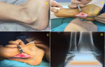
A 21-year-old male with six months of the instability of the ankle presented to our orthopedic department. He reported a trauma history in the ankle in which clinical and radiological findings were concluded to an ankle sprain and so a short leg was made along with oral´s analgesics and protected weight-bearing for two weeks. Following this, the pain had decreased but he reported recurrent episodes of a giving-away feeling of the ankle in sitting or kneeling. He could reproduce the dislocation by active dorsiflexion-eversion of his ankle. On the examination, he presented no deformity or wound. The palpation of the lateral side of the ankle was slightly painful. The subluxation of the peroneal tendons clinically appeared. The peroneal tendons dislocation test was positive. The X-rays of the ankle were normal. A few days later, the patient underwent surgery. The retinaculum was re-attached under the lip of the fibula by three anchors using the retromalleolar approach. After which, the ankle was placed in a below-knee, non-weight-bearing temporary cast for 2 weeks. Then walker boot cast with fully weight-bearing was allowed for 4 weeks. Following this period, physiotherapy was started. At the last follow up, the patient did well with neither instability nor ankle pain.
Figure 1: A) clinical appearance of dislocation of the peroneal tendons; B) intra-operative aspect showed the dislocation of the tendons; C) intra-operative aspect after the reattachment of the retinaculum; D) the post-operative x-ray showed the position of the anchors
Search
This article authors
On Pubmed
On Google Scholar
Citation [Download]
Navigate this article
Similar articles in
Key words
Article metrics

