
Infiltrating ocular dermolipoma leading to diplopia
Aymane Ridallah, Karima Madbouhi
Corresponding author: Aymane Ridallah, University Mohammed V Souissi, Ophtalmologie A, l´Hôpital des Spécialités, CHU Rabat, Rabat, Maroc 
Received: 02 Jun 2020 - Accepted: 24 Jul 2020 - Published: 26 Jul 2020
Domain: Ophthalmology
Keywords: Dermolipoma, diplopia, tumor
©Aymane Ridallah et al. PAMJ Clinical Medicine (ISSN: 2707-2797). This is an Open Access article distributed under the terms of the Creative Commons Attribution International 4.0 License (https://creativecommons.org/licenses/by/4.0/), which permits unrestricted use, distribution, and reproduction in any medium, provided the original work is properly cited.
Cite this article: Aymane Ridallah et al. Infiltrating ocular dermolipoma leading to diplopia. PAMJ Clinical Medicine. 2020;3:136. [doi: 10.11604/pamj-cm.2020.3.136.23968]
Available online at: https://www.clinical-medicine.panafrican-med-journal.com//content/article/3/136/full
Images in clinical medicine 

Infiltrating ocular dermolipoma leading to diplopia
Infiltrating ocular dermolipoma leading to diplopia
Aymane Ridallah1,&, Karima Madbouhi1
&Corresponding author

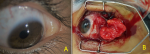
We report the observation of a 12-year-old child, who had left lower temporal swelling, gradually increasing in volume, evolving over several years, causing discomfort to ocular motility with binocular diplopia in the abduction look. The visual acuity was at 60/60 in both eyes, the anterior and posterior segment were without particularities. The examination found a voluminous «yellow» conjunctival swelling in inferior-temporal of left eye, not reaching the limb and whose size is unmodifiable. The eye motility examination found a limitation of abduction of the left eye, confirmed by a Lancaster test. An orbito-cerebral MRI was performed, revealing an orbital tumor of «greasy» density, with a suspicion of muscle infiltration, strongly suggestive of a dermolipoma. The patient had a surgical resection of the tumor, with a good post-operative evolution. The conjunctival dermolipoma is a benign congenital tumor, usually located at the temporal region of the bulbous conjunctiva, near the lateral canthus, it represents 5% of all orbital lesions in children. Dermolipoma are most often asymptomatic, or may progress during the first or second decade of life, and become visible in the primary eye or in adduction, requiring surgery for cosmetic purposes. However, many precautions must be taken, as cases of diplopia, strabismus, blepharoptosis and dry Keratocontivitis, were reported after surgery, following aggressive resection; by damaging the right muscles, the upper eyelid lifter and the tear gland. Therefore, resection of the dermolipoma should be treated more conservatively, to avoid this complications.
Figure 1: A) picture of the dermolipoma, hindering the left side look; B) intraoperative exposing of lipoma, after conjunctival resection
Search
This article authors
On Pubmed
On Google Scholar
Citation [Download]
Navigate this article
Similar articles in
Key words
Tables and figures
Article metrics
PlumX Metrics
Infiltrating ocular dermolipoma leading to diplopia
