
Bryan and Morrey Type IV intra-articular fracture: a rare situation
Ayoub Bouya, Omar Zaddoug
Corresponding author: Ayoub Bouya, Orthopedic Trauma Service I, Military Training Hospital Mohamed V, Rabat, Morocco 
Received: 14 Jun 2020 - Accepted: 24 Jul 2020 - Published: 27 Jul 2020
Domain: Orthopedic surgery
Keywords: Bryan and Morrey classification; coronal fracture, elbow
©Ayoub Bouya et al. PAMJ Clinical Medicine (ISSN: 2707-2797). This is an Open Access article distributed under the terms of the Creative Commons Attribution International 4.0 License (https://creativecommons.org/licenses/by/4.0/), which permits unrestricted use, distribution, and reproduction in any medium, provided the original work is properly cited.
Cite this article: Ayoub Bouya et al. Bryan and Morrey Type IV intra-articular fracture: a rare situation. PAMJ Clinical Medicine. 2020;3:138. [doi: 10.11604/pamj-cm.2020.3.138.24309]
Available online at: https://www.clinical-medicine.panafrican-med-journal.com//content/article/3/138/full
Images in clinical medicine 

Bryan and Morrey Type IV intra-articular fracture: a rare situation
Bryan and Morrey Type IV intra-articular fracture: a rare situation
Ayoub Bouya1,&, Omar Zaddoug1
&Corresponding author

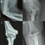
A 34-year-old man, a nurse with no medical history, non-smoking, and right-handed, admitted to the emergency service after a right elbow trauma. He falls on an outstretched hand on his house stairs. The patient couldn´t move his joint. The examination found a swelling and painful right elbow. Motion attempts were remarkably painful. Bony landmarks of the elbow were normal. There were no vascular or nervous abnormalities. The right wrist and shoulder exam were satisfying. Elbow X-rays revealed an articular displaced fracture of the distal humerus. 3D reconstruction affirmed the fracture. This frontal fracture takes the capitulum and the hole trochlea; It´s a was a Bryan and Morrey Type IV intra-articular fracture. Due to the displacement of this articular fracture, the staff decided to operate the patient. Within 24 hours after the trauma, the patient was in the operating room. Under loco-regional anesthesia, per operative examination of lateral ligaments was normal. An orthopedic surgeon performed the gesture by a combined approach (lateral and medial). The fragment was reduced and fixed par two Herbert screws. The articular congruity was satisfying. After three weeks of immobilization by a posterior splint, the patient debuted functional rehabilitation who been initially passive than active based on flexion-extension motion and pronation-supination motion. At 14 months following up, the patient had no pain. Ranges of motion were 150° of flexion, -10° of extension, 90° of pronation, and 80° of supination. Morrey score was 95. The limitation of extension motion had no impact on the usual activities of patient life.
Figure 1: Bryan and Morrey Type IV intra-articular fracture: a rare situation
Search
This article authors
On Pubmed
On Google Scholar
Citation [Download]
Navigate this article
Similar articles in
Key words
Article metrics

