
The demodex: the non-incriminated suspect of chronic blepharitis
Aymane Ridallah, Lalla Ouafaa Cherkaoui
Corresponding author: Aymane Ridallah, University Mohammed V Souissi, Ophtalmologie A, l´Hôpital des Spécialités, Centre Hospitalier Universitaire de Rabat, Rabat, Maroc 
Received: 15 Jun 2020 - Accepted: 24 Jul 2020 - Published: 30 Jul 2020
Domain: Ophthalmology
Keywords: Demodex folliculorum, Demodex brevis, blepharitis, MGD
©Aymane Ridallah et al. PAMJ Clinical Medicine (ISSN: 2707-2797). This is an Open Access article distributed under the terms of the Creative Commons Attribution International 4.0 License (https://creativecommons.org/licenses/by/4.0/), which permits unrestricted use, distribution, and reproduction in any medium, provided the original work is properly cited.
Cite this article: Aymane Ridallah et al. The demodex: the non-incriminated suspect of chronic blepharitis. PAMJ Clinical Medicine. 2020;3:149. [doi: 10.11604/pamj-cm.2020.3.149.24335]
Available online at: https://www.clinical-medicine.panafrican-med-journal.com//content/article/3/149/full
Images in clinical medicine 

The demodex: the non-incriminated suspect of chronic blepharitis
The demodex: the non-incriminated suspect of chronic blepharitis
Aymane Ridallah1,&, Lalla Ouafaa Cherkaoui1
&Corresponding author

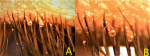
We report the case of a 55-year-old patient, followed in ophthalmology for recurrent chronic blepharitis for 5 years. The ophthalmological examination found a 20/20 visual acuity in both eyes, conjunctival hyperemia, instability of the tear film with a but less than 5 seconds, as well as the presence of a crusting anterior blepharitis, mainly located at the level of the upper eyelid, with microscopic evidence of cylindrical dandruff at the base of the eyelashes, corresponding to demodex. The patient was treated for blepharitis, including multiday palpebral hygiene, based on palpebral massage by hot compresses and combinations of eye-drops and ointments based on antibiotics and corticosteroids, all associated with an additional treatment with artificial tears. Eye demodex is a common condition, but clinically under-diagnosed; demodex mites have been incriminated in the physiopathology of anterior blepharitis (Demodex folliculorum) and posterior blepharitis (Demodex brevis). The folliculorum finds its main habitat at the base of the eyelash follicle, where it feeds on epithelial cells, causing direct mechanical damage. Since demodex does not have excretory organs, the undigested residues are regurgitated, and then combine with epithelial cells, keratin and eggs to form the cylindrical deposits of the eyelashes, characteristic of the presence of demodex. (Demodex brevis) was associated with dysfunction of the Meibomus glands, where it causes a primary mechanical blockage of the glandular orifice.
Figure 1: A) the patient´s upper eyelid, under microscopy, showing the presence of demodex at the base of the lashes (cylindrical dandruff); B) high magnification aspect
Search
This article authors
On Pubmed
On Google Scholar
Citation [Download]
Navigate this article
Similar articles in
Key words
Tables and figures
Article metrics

