
Successful thrombolysis in acute coronary syndrome
Najlaa Salmi, Safae Hilal
Corresponding author: Najlaa Salmi, Cardiology Department of the Military Hospital of Instruction Mohamed V of Rabat, Rabat, Morocco 
Received: 06 May 2020 - Accepted: 18 May 2020 - Published: 20 May 2020
Domain: Cardiology
Keywords: Acute coronary syndrome, thrombolysis, ECG
©Najlaa Salmi et al. PAMJ Clinical Medicine (ISSN: 2707-2797). This is an Open Access article distributed under the terms of the Creative Commons Attribution International 4.0 License (https://creativecommons.org/licenses/by/4.0/), which permits unrestricted use, distribution, and reproduction in any medium, provided the original work is properly cited.
Cite this article: Najlaa Salmi et al. Successful thrombolysis in acute coronary syndrome. PAMJ Clinical Medicine. 2020;3:23. [doi: 10.11604/pamj-cm.2020.3.23.23351]
Available online at: https://www.clinical-medicine.panafrican-med-journal.com//content/article/3/23/full
Images in clinical medicine 

Successful thrombolysis in acute coronary syndrome
Successful thrombolysis in acute coronary syndrome
Najlaa Salmi1,&, Safae Hilal1
1Cardiology Department of the Military Hospital of Instruction Mohamed V of Rabat, Rabat, Morocco
&Corresponding author
Najlaa Salmi, Cardiology Department of the Military Hospital of Instruction Mohamed V of Rabat, Rabat, Morocco

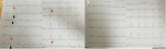
We report the case of a male aged by 50 years-old without medical history who consult in emergency for a prolonged constrictive thoracic left pain which irradiates to the left shoulder from 5 hours. Cardiologic and general examination were normal. In front of that we perform an ECG, who show a reciprocal ST-segment elevation at the inferior territory (DII, DIII, aVF), on anterior and lateral derivations (A). After the elimination of thrombolysis contraindications, we administrate first a loading dose of clopidogrel, aspirin, and enoxaparin followed by tenecteplase at 0.6. The immediate follow-up was marked by the resolution of the pain and a complete resolution of the ST-segment elevation synonym of the successful thrombolysis (B). Thrombolysis is a medical technique intended to urgently dissolve a curd that blurs a coronary artery in myocardial infarction or a cerebral artery in a stroke which success only in 15% of cases. Thrombolysis shows its effectiveness in terms of survival and improvement of left ventricular function. It´s indicated if the chest pain dates from less than 12 hours in the absence of contraindications. Its prescription methods consist of a single intravenous bolus injection, dosage depending on weight associated with treatment with aspirin, clopidogrel and effective anticoagulation. The success criteria are clinically: pain sedation after an initial increase possible, biologically an early peak in markers of myocardial necrosis, electrically ventricular reperfusion arrhythmia (ESV, TV, RIVA) and the marked regression see the disappearance of the ST segment elevation.
Figure 1: before thrombolysis ECG (A) show a reciprocal ST-segment elevation at the inferior territory (DII, DIII, aVF) (black arrows), on anterior and lateral derivations (red arrows). After thrombolysis ECG (B) show the complete resolution of the ST-segment elevation synonym of the success
Search
This article authors
On Pubmed
On Google Scholar
Citation [Download]
Navigate this article
Similar articles in
Key words
Article metrics
PlumX Metrics
Successful thrombolysis in acute coronary syndrome
