
Azygos lobe
Asaad El Bakkari, Ouadie El Menaoui
Corresponding author: Asaad El Bakkari, Radiology Department Military Hospital Mohamed Fifth of Instruction of Rabat Morocco 
Received: 24 Apr 2020 - Accepted: 27 May 2020 - Published: 28 May 2020
Domain: Radiology
Keywords: Azygos lobe, azygos vein, CT
©Asaad El Bakkari et al. PAMJ Clinical Medicine (ISSN: 2707-2797). This is an Open Access article distributed under the terms of the Creative Commons Attribution International 4.0 License (https://creativecommons.org/licenses/by/4.0/), which permits unrestricted use, distribution, and reproduction in any medium, provided the original work is properly cited.
Cite this article: Asaad El Bakkari et al. Azygos lobe. PAMJ Clinical Medicine. 2020;3:31. [doi: 10.11604/pamj-cm.2020.3.31.23075]
Available online at: https://www.clinical-medicine.panafrican-med-journal.com//content/article/3/31/full
Images in clinical medicine 

Azygos lobe
Azygos lobe
Asaad El Bakkari1,&, Ouadie El Menaoui1
1Radiology Department, Military Hospital Mohamed Fifth of Instruction of Rabat, Rabat, Morocco
&Corresponding author
Asaad El Bakkari, Radiology Department, Military Hospital Mohamed Fifth of Instruction of Rabat, Rabat, Morocco

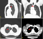
We report the case of a 25-year-old male who consult for a chronic dry cough without fever or medical history, in front of that we perform a chest X-ray completed by a chest-CT who shows a thin curvilinear density seen in the upper right lung, convex towards the chest wall at its base is the azygos vein which can be seen as a teardrop-shaped structure (A,B,C,D). The azygos lobe or fissure is a rare anatomic variant affecting about 1% of the general population. He is a summery lobe without pathological consequence on the breathing function, who form when posterior cardinal vein penetrates the upper medial portion of the apical segment of the right upper lobe, instead of remaining along the apex of the lung. This demarcating fissure contains both visceral and parietal pleural layers and has no bronchus, so is not a true accessory lobe with a variable size. The diagnosis is made incidentally and easily by chest X-ray or CT and does not require any treatment.
Figure 1: (A) sagittal section-CT; (B) coronal section-CT; (C) axial section-CT on pulmonar window; axial section-CT (D) on mediastinal window shows a thin curvilinear density seen in the upper right lung, convex towards the chest wall at its base is the azygos vein which can be seen as a teardrop-shaped structure (red arrows) which corresponds to an azygos fissure fissure
Search
This article authors
On Pubmed
On Google Scholar
Citation [Download]
Navigate this article
Similar articles in
Key words
Article metrics
PlumX Metrics
Azygos lobe
