
Cardiac tamponade by hydatid pericardial cyst: a rare echocardiography image
Amine Ech-chenbouli, Saoussane Serbout
Corresponding author: Amine Ech-chenbouli, Department of Cardiology Ibn Rochd University Hospital Casablanca, Casablanca, Morocco 
Received: 15 May 2020 - Accepted: 18 May 2020 - Published: 04 Jun 2020
Domain: Cardiology
Keywords: Hydatid cyst, tamponade, echocardiography
©Amine Ech-chenbouli et al. PAMJ Clinical Medicine (ISSN: 2707-2797). This is an Open Access article distributed under the terms of the Creative Commons Attribution International 4.0 License (https://creativecommons.org/licenses/by/4.0/), which permits unrestricted use, distribution, and reproduction in any medium, provided the original work is properly cited.
Cite this article: Amine Ech-chenbouli et al. Cardiac tamponade by hydatid pericardial cyst: a rare echocardiography image. PAMJ Clinical Medicine. 2020;3:39. [doi: 10.11604/pamj-cm.2020.3.39.23556]
Available online at: https://www.clinical-medicine.panafrican-med-journal.com//content/article/3/39/full
Images in clinical medicine 

Cardiac tamponade by hydatid pericardial cyst: a rare echocardiography image
Cardiac tamponade by hydatid pericardial cyst: a rare echocardiography image
Amine Ech-chenbouli 1,&, Saoussane Serbout1
1Department of Cardiology Ibn Rochd University Hospital Casablanca, Casablanca, Morocco
&Corresponding author
Amine Ech-chenbouli, Department of Cardiology Ibn Rochd University Hospital Casablanca,
Casablanca, Morocco

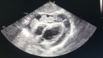
A 19-year-old previously healthy woman originaly from the countryside of Morocco presented to our emergency department with dyspnea chest discomfort and hypotension. She reported a 1-month history of fever and abdominal pain. On physical examination her heart sounds were rhythmic but muffled, and there was an audible pericardial friction rub. Her EKG showed sinus tachycardia with low voltage. Two-dimensional transthoracic echocardiography revealed large pericardial effusion with the presence of round formations in the pericardial space suggestive of hydatid cysts. Abdominal CT showed rupture of a hepatic hydatid cyst into the pericardial space through a transdiaphragmatic fistula. Diagnostic of Cardiac tamponade by hydatid pericardial cyst: was confirmed. The patient underwent urgent surgical drainage of her pericardial effusion and was medically treated with albendazole. Unfortunately she died 2 days after her surgery.
Figure 1: a subcostal 4 chamber transthoracic echocardiographic view showing an important pericardial effusion with the presence of hydatid cysts
Search
This article authors
On Pubmed
On Google Scholar
Citation [Download]
Navigate this article
Similar articles in
Key words
Tables and figures
Article metrics

