
A huge idiopathic pulmonary aneurysm: a stable follow up
Meryem El Ghanmi, Jamal Fatihi
Corresponding author: Meryem El Ghanmi, Cardiology Department , Mohamed V Military Hospital of Rabat, Rabat, Morocco 
Received: 25 May 2020 - Accepted: 29 May 2020 - Published: 10 Jun 2020
Domain: Cardiology
Keywords: Pulmonary artery , aneurysm , CT- scann
©Meryem El Ghanmi et al. PAMJ Clinical Medicine (ISSN: 2707-2797). This is an Open Access article distributed under the terms of the Creative Commons Attribution International 4.0 License (https://creativecommons.org/licenses/by/4.0/), which permits unrestricted use, distribution, and reproduction in any medium, provided the original work is properly cited.
Cite this article: Meryem El Ghanmi et al. A huge idiopathic pulmonary aneurysm: a stable follow up. PAMJ Clinical Medicine. 2020;3:49. [doi: 10.11604/pamj-cm.2020.3.49.23738]
Available online at: https://www.clinical-medicine.panafrican-med-journal.com//content/article/3/49/full
Images in clinical medicine 

A huge idiopathic pulmonary aneurysm: a stable follow up
A huge idiopathic pulmonary aneurysm: a stable follow up
Meryem El Ghanmi1,&, Jamal Fatihi2
1Cardiology Department , Mohamed V Military Hospital of Rabat, Rabat, Morocco, 2Internal Medicine Department, Mohamed V Military Hospital of Rabat, Rabat, Morocco
&Corresponding author
Meryem El Ghanmi, Cardiology Department , Mohamed V Military Hospital of Rabat, Rabat, Morocco

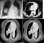
A 60 years old woman, presented 5 years ago to the cardiology department addressed by her physician for atypical chest pain. She reported no history of palpitation or dyspnea. The cardiac auscultation revealed a 3/6 systolic murmur at the left lower sternal border. The Chest radiography showed cardiomegaly with homogenous bilateral hilar opacities (A). The chest computed tomography (CT) angiography revealed a huge aneurysm of the main pulmonary artery sized 105 mm in transverse diameter (B,C,D) extending up to both left and right pulmonary arteries. It was diagnosed as idiopathic because the patient had no comorbidities that can cause a pulmonary artery (PA) aneurysm. We recommended surgery for the PA aneurysm, but the patient refused surgery, she was discharged with medication and regular follow-up; her clinical condition stayed stable and the CT scan annually controls didn't show any increase in the PA diameter.
Figure 1: (A) chest radiography with a bilateral hilar opacities; (B,C,D) CT-scan showing the aneurysm of the main PA and its branches
Search
This article authors
On Pubmed
On Google Scholar
Citation [Download]
Navigate this article
Similar articles in
Key words
Tables and figures
Article metrics
PlumX Metrics
A huge idiopathic pulmonary aneurysm: a stable follow up
