
Luxation postérieure bilatérale spontanée du cristallin
Narjisse Taouri, Ouafae Cherkaoui
Corresponding author: Narjisse Taouri, Department A of Ophthalmology, Mohammed V University Souissi, Rabat, Morocco 
Received: 07 May 2020 - Accepted: 18 May 2020 - Published: 24 Jun 2020
Domain: Ophthalmology
Keywords: Syndrome pseudoexfoliatif, microfibrillopathie élastique, instabilité zonulaire, luxation cristallin
©Narjisse Taouri et al. PAMJ Clinical Medicine (ISSN: 2707-2797). This is an Open Access article distributed under the terms of the Creative Commons Attribution International 4.0 License (https://creativecommons.org/licenses/by/4.0/), which permits unrestricted use, distribution, and reproduction in any medium, provided the original work is properly cited.
Cite this article: Narjisse Taouri et al. Luxation postérieure bilatérale spontanée du cristallin. PAMJ Clinical Medicine. 2020;3:71. [doi: 10.11604/pamj-cm.2020.3.71.23373]
Available online at: https://www.clinical-medicine.panafrican-med-journal.com//content/article/3/71/full
Images in clinical medicine 

Luxation postérieure bilatérale spontanée du cristallin
Luxation postérieure bilatérale spontanée du cristallin
Spontaneous bilateral posterior dislocation of the crystalline lens
Narjisse Taouri1,&, Ouafae Cherkaoui1
1Department A of Ophthalmology, Mohammed V University Souissi, Rabat, Morocco
&Auteur correspondant
Narjisse Taouri, Department A of Ophthalmology, Mohammed V University Souissi, Rabat, Morocco
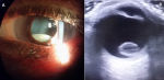
We report the case of an 89-year old female patient with a history of high blood pressure without ocular trauma. Ophthalmological examination showed bilateral visual acuity of counting fingers. Bilaterally, biomicroscopy found clear cornea, good anterior chamber, exfoliation material deposit at the edge of the pupil. In the left eye, crystalline lens was posteriorly subluxated but still visible in the pupillary area (A). Right eye fundus examination revealed dislocation of the crystalline lens into the vitreous (B). The diagnosis of spontaneous bilateral dislocation of the crystalline lens secondary to pseudoexfoliation syndrome was established. Data from the literature highlight that pseudoexfoliation syndrome is an age-related systemic disease associated with elastic microfibrillopathy due to altered LOXL1 gene. This results in the production and accumulation of fibrillar deposits within the extracellular matrix of several organs. This syndrome is most often bilateral and asymmetric. Clinical manifestations include cataract, glaucoma, bad pupillary dilatation, phacodonesis or spontaneous subluxation of the crystalline lens into the vitreous due to zonular instability. It has been reported in the literature that this zonular instability may be due to disruption secondary to the deposit of fibrillary material at the pre-equatorial zonule insertion. Other studies have reported that this may be intrinsic, given that, in affected eyes, zonular fiber examination shows differences in elastic fibers with respect to unaffected eyes.
Key words: Pseudoexfoliation syndrome, elastic microfibrillopathy, zonular instability, crystalline lens dislocation

Nous rapportons le cas d´une patiente âgée de 89 ans, ayant comme antécédent de l´hypertension artérielle, sans notion de traumatisme oculaire, chez qui l´examen ophtalmologique a retrouvé une acuité visuelle à compte les doigts bilatéralement. Et l´examen biomicroscopique a retrouvé une cornée claire, une bonne chambre antérieure, dépôt de matériel exfoliatif sur le bord pupillaire bilatéralement. Au niveau de l´œil gauche le cristallin était subluxé en postérieur et encore visible dans l´aire pupillaire (A). Et au niveau de l´œil droit, l´examen du fond d´œil a mis en évidence le cristallin luxé dans le vitré (B). Le diagnostic était une luxation spontanée bilatérale du cristallin secondaire au syndrome pseudoexfoliatif. Selon les données de la littérature, le syndrome pseudoexfoliatif est une maladie systémique liée à l´âge, en rapport avec une microfibrillopathie élastique suite à une altération du gène Lysyl oxidase homolog 1 (LOXL1). Et donc comme conséquence on a une production et une accumulation de dépôts fibrillaires au sein de la matrice extracellulaire de plusieurs organes. Au niveau oculaire, l`atteinte souvent bilatérale et asymétrique, ce syndrome peut se manifeste cliniquement par : cataracte, glaucome, la mauvaise dilatation pupillaire, phacodonésis ou subluxation spontanée du cristallin dans le vitré suite à une instabilité zonulaire. Des auteurs ont rapporté que cette instabilité zonulaire peut être due à une disruption secondaire au dépôt du matériel fibrillaire sur l´insertion pré-équatorial de la zonule. Et d´autres ont rapporté que ça peut être d´origine intrinsèque, vu que l´étude des fibres zonulaires des yeux atteints a retrouvé des fibres élastiques différentes par rapport aux yeux non atteints.
Figure 1 : A) œil
gauche : équateur du cristallin visible dans la pupille ; B) œil
droit : luxation postérieure du cristallin vue sur échographie
en mode B
Search
This article authors
On Pubmed
On Google Scholar
Citation [Download]
Navigate this article
Similar articles in
Key words
Tables and figures
Article metrics
PlumX Metrics
Luxation postérieure bilatérale spontanée du cristallin
