
Cervical cystic lymphangioma
Zakia El Yousfi, Najwa Ech-Cherif El Kettani
Corresponding author: Zakia El Yousfi,Neuroradiology Department Head and Neck Hospital of Rabat, Rabat, Morocco 
Received: 31 May 2020 - Accepted: 27 Jun 2020 - Published: 08 Jul 2020
Domain: Radiology,Otolaryngology (ENT)
Keywords: Lymphangioma, cervical, CT
©Zakia El Yousfi et al. PAMJ Clinical Medicine (ISSN: 2707-2797). This is an Open Access article distributed under the terms of the Creative Commons Attribution International 4.0 License (https://creativecommons.org/licenses/by/4.0/), which permits unrestricted use, distribution, and reproduction in any medium, provided the original work is properly cited.
Cite this article: Zakia El Yousfi et al. Cervical cystic lymphangioma. PAMJ Clinical Medicine. 2020;3:94. [doi: 10.11604/pamj-cm.2020.3.94.23879]
Available online at: https://www.clinical-medicine.panafrican-med-journal.com//content/article/3/94/full
Images in clinical medicine 

Cervical cystic lymphangioma
Cervical cystic lymphangioma
Zakia El Yousfi1,&, Najwa Ech-Cherif El Kettani1
&Corresponding author

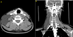
Our work is about a 34-year-old woman with no particular history who was admitted for a cervical mass which showed a substantial increase in size in the last 8 months. Physical examination revealed a fluctuant and painless mass in the right posterior triangle which was tender on palpation. CT scan of the neck showed a multi-loculated homogeneous cystic lesion located primarily in the right posterior cervical triangle, and extending to the right deep cervical spaces, underneath an elevated sternocleidomastoid muscle. The right jugular vein, while relatively compressed, was still patent. There was no contrast enhancement after intravenous injection. The patient underwent complete surgical excision by transverse neck incision and the diagnosis of lymphangioma was confirmed on histological examination. Cystic lymphangiomas are rare tumors of variable locations and their cause remains unknown. There is no racial or gender predilection. The most common site for the tumor is the posterior triangle of the neck. The clinical picture allows the suspicion of lymphangioma, CT more precisely determines the relationship of the mass with adjacent structures. However, the definitive diagnosis is only obtained with the final pathology.
Figure 1: axial CT section (A) and coronal CT section (B) shows a multi-loculated cystic lesion in the right posterior cervical triangle extending to the right deep cervical spaces
Search
This article authors
On Pubmed
On Google Scholar
Citation [Download]
Navigate this article
Similar articles in
Key words
Article metrics
PlumX Metrics
Cervical cystic lymphangioma
