Syndrome de Sturg-Weber
Karmoun Souhaila, Berraho Amina
Corresponding author: Karmoun Souhaila, Service d´Ophtalmologie B, Hôpital des Spécialités, Rabat, Maroc 
Received: 08 Jun 2020 - Accepted: 24 Jul 2020 - Published: 13 Oct 2020
Domain: Ophthalmology
Keywords: Syndrome de Sturge-Weber, glaucome, angiome hémifacial
©Karmoun Souhaila et al. PAMJ Clinical Medicine (ISSN: 2707-2797). This is an Open Access article distributed under the terms of the Creative Commons Attribution International 4.0 License (https://creativecommons.org/licenses/by/4.0/), which permits unrestricted use, distribution, and reproduction in any medium, provided the original work is properly cited.
Cite this article: Karmoun Souhaila et al. Syndrome de Sturg-Weber. PAMJ Clinical Medicine. 2020;4:63. [doi: 10.11604/pamj-cm.2020.4.63.24158]
Available online at: https://www.clinical-medicine.panafrican-med-journal.com//content/article/4/63/full
Images in clinical medicine 

Syndrome de Sturg-Weber
Syndrome de Sturg-Weber
Sturge-Weber syndrome
Karmoun Souhaila1,&, Berraho Amina1
&Auteur correspondant
We report a case of a 3-year-old child, presented to the ophthalmic consultation by his parents. They noticed divergent strabismus in the child´s right eye associated with tearing. In examination, a notion of first-degree parental consanguinity was revealed. The child has an angioma of the right hemiface. The patient underwent an ophthalmological examination with general anesthesia which objectified in the right eye a corneal diameter at 14mm, a clear cornea, a deep anterior chamber, and a papillary excavation at 8/10 in the fundus. The examination of the left eye finds a corneal diameter at 12mm, a clear cornea and a papillary excavation at 6/10 in the fundus. The eye pression at Perkins was 22mmhg and 14mmhg for the right and left eye respectively. Sturge-Weber syndrome was retained as a positive diagnosis. Sturge-Weber syndrome is a rare congenital encephalotrigeminal angiomatosis which, in its complete form, associates a facial angioma that always occupies the territory of V1, even if it can sometimes invade a larger facial area, and ocular (glaucoma, choroidal angioma) also homolateral. Glaucoma secondary to this syndrome can be severe due to its early onset and resistance to conventional therapy.
Key words: Sturge-Weber syndrome, glaucoma, hemifacial angioma

Nous rapportons le cas d´un enfant âgé de 3 ans, sans aucuns antécédents notables, mené à la consultation ophtalmologique devant la constatation des parents d´un strabisme divergent au niveau de l´œil droit associé à un larmoiement. L´interrogatoire minutieux a dévoilé une notion de consanguinité parentale du premier degré. L´enfant présente un angiome de l´hémiface droite. Le patient a bénéficié d´un examen ophtalmologique sous anesthésie générale qui a objectivé en œil droit ayant un diamètre cornéen à 14 mm, une cornée claire, une chambre antérieure profonde et au fond d´œil une excavation papillaire à 8/10. L´examen au niveau de l´œil gauche trouve un diamètre cornéen à 12 mm, une cornée claire et au fond d´œil une excavation papillaire à 6/10. Le tonus oculaire au Perkins était respectivement à 22mmhg et 14mmhg pour l´œil droit et gauche. Le syndrome de Sturge-Weber a été retenu comme diagnostic positif. Le syndrome de Sturge-Weber est une angiomatose encéphalo-trigéminée congénitale rare qui, dans sa forme complète, associe un angiome plan facial qui occupe toujours le territoire du V1, même s'il peut parfois envahir une plus grande aire faciale et une atteinte oculaire (glaucome, angiome choroïdien) également homolatérale. Le glaucome secondaire à ce syndrome peut être grave en raison de son apparition précoce et résistance à la thérapie conventionnelle.
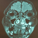
Figure 1: (A,B) mégalocornée au niveau de l'œil droit et angiome de l'hémiface homolatéral
Search

This article authors
On Pubmed
On Google Scholar
Citation [Download]
Navigate this article
Similar articles in
Key words
Tables and figures

Article metrics
PlumX Metrics
Syndrome de Sturg-WeberRecently from the PAMJ-CM