
Vegetating cutaneous plaques: a case report
Selma El Kadiri, Zakia Douhi, Sara Elloudi, Hanane Baybay, Fatima-Zahra Mernissi, Amal Douida, Layla Tahiri, Nawal Hammas, Leila Chbani, Hinde El Fatemi
Corresponding author: Selma El Kadiri, Department of Dermatology, CHU Hassan II, Fez, Morocco 
Received: 25 Dec 2020 - Accepted: 18 Jan 2021 - Published: 19 Jan 2021
Domain: Dermatology
Keywords: Pemphigus vegetans, seborrheic keratosis like, rituximab
©Selma El Kadiri et al. PAMJ Clinical Medicine (ISSN: 2707-2797). This is an Open Access article distributed under the terms of the Creative Commons Attribution International 4.0 License (https://creativecommons.org/licenses/by/4.0/), which permits unrestricted use, distribution, and reproduction in any medium, provided the original work is properly cited.
Cite this article: Selma El Kadiri et al. Vegetating cutaneous plaques: a case report. PAMJ Clinical Medicine. 2021;5:19. [doi: 10.11604/pamj-cm.2021.5.19.27583]
Available online at: https://www.clinical-medicine.panafrican-med-journal.com//content/article/5/19/full
Case report 

Vegetating cutaneous plaques: a case report
Vegetating cutaneous plaques: a case report
Selma El Kadiri1,&, Zakia Douhi1, Sara Elloudi1, Hanane Baybay1, Fatima-Zahra Mernissi1, Amal Douida2, Layla Tahiri2, Nawal Hammas2, Leila Chbani2, Hinde El Fatemi2
&Corresponding author
Pemphigus vegetans is a rare variant of pemphigus vulgaris that manifests as localized plaques in two classic subtypes, the Neumann type, which resembles pemphigus vulgaris with initial vesicular and erosive lesions; the Hallopeau type with pustular lesions and which usually remains localized. Here we report a case of a 44-year-old patient admitted for itchy lesions over the inframammary fold, groin, perianal area and limbs. Initially, they were flaccid blisters that became crusted erosions for the past 6 months evolving finally into vegetating plaques. Histological examination showed acanthosis, papillomatosis, acantholysis and the presence of microabscesses containing eosinophils. Direct immunofluorescence showed intercellular immunoglobulin G (IgG) deposits in the epidermis. The diagnosis of pemphigus vegetans Neumann type was confirmed. She was treated with an oral corticosteroid (0.5mg/kg per day) and rituximab, an anti-CD20 monoclonal antibody 1g 15 days apart with significant regression of the lesions.

Pemphigus vegetans represent only 1-2% of all cases of pemphigus [1]. It has two subtypes: Neumann and Hallopeau. The Neumann form initially begins with vesicular, flaccid bullae and erosive lesions resembling to a typical pemphigus vulgaris. Contrary to the less common Hallopeau type that starts with pustules and remains localized with a relatively benign course [2]. The disease affects the axilla, inframammary, intergluteal and inguinal folds inciting formation of vegetations maintained by maceration, friction and infection [3]. We report here a case of pemphigus vegetans with exclusive cutaneous vegetations.

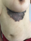
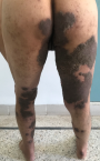
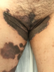
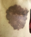
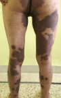
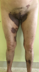
A 44-year-old Moroccan female hospitalized for itchy lesions over inframammary fold, groin, perianal area and limbs. The skin lesions started with flaccid blisters that become crusted erosions for the past 6 months. They finally evolved into vegetating plaques. She denied systemic symptoms or the ingestion of suspicious drugs. The patient was previously treated by antibiotics without improvement. Laboratory tests were carried out and revealed diabetes put on metformin and glimepiride. Clinical examination revealed well-defined, multiple dark brown, indurated, superelevated plaques with verrucous and seborrheic-keratosis like surface over the infra-mammary area (Figure 1), back, both legs, genital and perianal area (Figure 2, Figure 3).
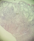
Nikolsky's sign was negative. Buccal, genital, nasal and ocular mucosae were spared. General physical and systemic examination revealed nothing abnormal. Complete blood count revealed peripheral eosinophilia. Histological examination showed acanthosis, papillomatosis, acantholysis and the presence of microabscesses containing eosinophils (Figure 4). Direct immunofluorescence showed intercellular immunoglobulin G (IgG) deposits in the epidermis. Indirect immunofluorescence revealed IgG autoantibodies directed against desmosomes. The diagnosis of pemphigus vegetans Neumann type was confirmed. She was treated with oral corticosteroid (0.5mg/kg per day) and rituximab, an anti-CD20 monoclonal antibody 1g 15 days apart with significant regression of the lesions (Figure 5, Figure 6, Figure 7).
Pemphigus vegetans is a rare disease that may begin with flaccid blisters that become erosions and form papillomatous proliferations localized especially in folds or on the scalp [1]. The tongue often shows cerebriform morphologic features early in the course of the disease [2]. Verrucous plaques on the scalp, fingers, perianal and genital areas have been reported as the mode of onset of the disease [3]. The lesions evolve to coalesce forming large patches into groups or figurate patterns [3]. In our case, the patient didn´t have mucosal or scalp involvement.
Our patient had lesions over the flexures and extensors with no involvement of the mucosa. She had a pattern of sulci, gyri over her lesions corresponding to a cerebriform pattern which may probably correspond to the papillary hyperplasia as reported once before by Rebello et al. [4]. Cases induced by the ingestion of the drugs such as captopril and enalapril and by the intranasal use of heroin and cocaine have been reported. The association with HIV infection and gastric cancer may have been fortuitous [3].
Pemphigus vegetans must be differentiated from other causes of vegetations such as halogenoderma which includes ioderma, bromoderma and fluoroderma, blastomycosis-like pyoderma, chromoblastomycosis, pyodermatitis-pyostomatitis vegetans, granuloma inguinal, condyloma lata and amebic granulomas specially when there is no involvement of the mucosa [3]. The laboratory findings, epidemiology, pathogenesis and treatment of pemphigus vegetans are the same as those for pemphigus vulgaris [4]. Desmocollin-3 antibodies have been reported in some cases and hypothesized to explain the different clinical features in vegetans versus vulgaris [5].
We report a cerebriform pattern of cutaneous pemphigus vegetans with sulci and gyri sparing mucosa. So in routine pratices, extensive cerebriform lesions over the flexures and extensors too and also pemphigus vegetans should be kept as good differential.
The authors declare no competing interests.
All the authors have read and agreed to the final manuscript.
Figure 1: verrucous and vegetating plaques over inframammary area
Figure 2: vegetative hyperkeratotic plaques in limbs and perianal area
Figure 3: perineal involvement with vegetative hyperkeratotic plaques in limbs
Figure 4: histological examination showed acanthosis, papillomatosis, acantholysis and the presence of microabscesses containing eosinophils
Figure 5: re-examination 3 months later showing clinical remission in the inframammary area
Figure 6: re-examination 3 months later showing clinical remission in the perianal area and the limbs
Figure 7: re-examination 3 months later showing clinical remission in the genital area and the limbs
- Ahmed AR, Blose DA. Pemphigus vegetans: Neumann type and Hallopeau type. Int J Dermatol. 1984;23(2):135-41. PubMed | Google Scholar
- Premalatha S, Jayakumar S, Yesudian P, Thambiah AS. Cerebriform tongue: a clinical sign in pemphigus vegetans. Br J Dermatol. 1981;104(5):587-91. PubMed | Google Scholar
- Verma GK, Tegta GR, Sharma A, Kaur M, Sharma S. A rare case of extensive pemphigus vegetans. Indian Dermatology Online Journal. 2020 Jan-Feb;11(1):87-90. PubMed | Google Scholar
- Rebello MS, Ramesh BM, Sukumar D, Alapatt GF. Cerebriform cutaneous lesions in pemphigus vegetans. Indian J Dermatol. 2016;61(2):206-8. PubMed | Google Scholar
- Saruta H, Ishii N, Teye K, Ono F, Ohyama B, Koga H et al. Two cases of pemphigus vegetans with IgG anti-desmocollin 3 antibodies. JAMA Dermatol. 2013;149(10):1209-13. PubMed | Google Scholar
Search
This article authors
On Pubmed
On Google Scholar
Citation [Download]
Navigate this article
Similar articles in
Key words
Tables and figures
Article metrics
PlumX Metrics
Vegetating cutaneous plaques: a case report
