
Deep venous thrombosis with multifocal tuberculosis in an immunocompetent patient
Inas El Kacemi, Brahim El Mostarchid
Corresponding author: Inas El Kacemi, Service de Neurochirurgie, Faculté de Médecine et de Pharmacie, l´Hôpital Militaire d´Instruction Mohammed V-Rabat, Université Mohammed V de Rabat, Rabat, Maroc 
Received: 07 Nov 2020 - Accepted: 18 Jan 2021 - Published: 19 Jan 2021
Domain: Neurosurgery
Keywords: Deep venous thrombosis, multifocal tuberculosis, immunocompetent patient
©Inas El Kacemi et al. PAMJ Clinical Medicine (ISSN: 2707-2797). This is an Open Access article distributed under the terms of the Creative Commons Attribution International 4.0 License (https://creativecommons.org/licenses/by/4.0/), which permits unrestricted use, distribution, and reproduction in any medium, provided the original work is properly cited.
Cite this article: Inas El Kacemi et al. Deep venous thrombosis with multifocal tuberculosis in an immunocompetent patient. PAMJ Clinical Medicine. 2021;5:22. [doi: 10.11604/pamj-cm.2021.5.22.26861]
Available online at: https://www.clinical-medicine.panafrican-med-journal.com//content/article/5/22/full
Images in clinical medicine 

Deep venous thrombosis with multifocal tuberculosis in an immunocompetent patient
Deep venous thrombosis with multifocal tuberculosis in an immunocompetent patient
Inas El Kacemi1,&, Brahim El Mostarchid1
&Corresponding author

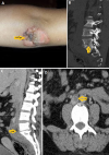
A 33-year-old woman with no notable history who consults for a tuberculous gumma lesion in the elbow dating back 1 month with concurrent concomitant low back pain and all this in a context of night sweats and unencrypted weight loss. The cutaneous biopsy revealed granulomas with absence of caseous necrosis. The radiological assessment based on lumbar CT scan showed L5-S1 mirror images with an anterior vertebral collection engulfing the large retroperitoneal vessels and the angio-scan confirmed the thrombosis of the inferior vena cava, the scanned-guided biopsy confirmed the diagnosis of tuberculous spondylo-discitis. The remainder of the report showed a positive intradermal reaction (IDR) with negative VIH serology. The diagnosis of multifocal tuberculosis involving cutaneous, osteoarticular and pulmonary involvement in an immunocompetent patient was selected and the patient was placed on anti-TB drugs under the 2 months of streptomycin, rifampicine, isoniazid and pyrazinamide followed by rifampicine and isoniazid for 10 months, with good clinical evolution. The multifocal forms of tuberculosis represent 9 to 10% of cases and are the preserve of immunocompromised patients mainly carriers of VIH. Their prognosis is poor with a mortality rate of 16 to 25%. They also pose a real problem of diagnosis, given its clinical polymorphism and the diversity of organs that can be affected, which is the cause of the delay in diagnosis.
Figure 1: A) tuberculous gumma of the elbow; B) lumbar CT scan showed L5-S1 mirror images; C) lumbar CT scan showed L5-S1 anterior vertebral collection engulfing the large retroperitoneal vessels; D) angio-scan confirmed the thrombosis of the inferior vena cava
Search
This article authors
On Pubmed
On Google Scholar
Citation [Download]
Navigate this article
Similar articles in
Key words
Article metrics

