
Electrocardiogram (ECG) changes of a patient with severe hyperkalemia
Sarvesh Seger
Corresponding author: Sarvesh Seger, School of Medicine, International Medical University, Clinical Campus Kluang, Kluang, Johor, Malaysia 
Received: 01 Dec 2020 - Accepted: 08 Feb 2021 - Published: 09 Feb 2021
Domain: Emergency medicine,Internal medicine,Nephrology
Keywords: Hyperkalemia, electrocardiogram, emergency, Brugada, end stage renal disease
©Sarvesh Seger et al. PAMJ Clinical Medicine (ISSN: 2707-2797). This is an Open Access article distributed under the terms of the Creative Commons Attribution International 4.0 License (https://creativecommons.org/licenses/by/4.0/), which permits unrestricted use, distribution, and reproduction in any medium, provided the original work is properly cited.
Cite this article: Sarvesh Seger et al. Electrocardiogram (ECG) changes of a patient with severe hyperkalemia. PAMJ Clinical Medicine. 2021;5:55. [doi: 10.11604/pamj-cm.2021.5.55.27254]
Available online at: https://www.clinical-medicine.panafrican-med-journal.com//content/article/5/55/full
Images in clinical medicine 

Electrocardiogram (ECG) changes of a patient with severe hyperkalemia
Electrocardiogram (ECG) changes of a patient with severe hyperkalemia
Sarvesh Seger1,&
&Corresponding author

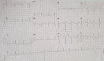
A 55-year-old gentleman with underlying type 2 diabetes mellitus, dyslipidemia, hypertension, and end stage renal disease presented with shortness of breath, vomiting and worsening fatigue for one day after missing his scheduled hemodialysis. Physical examination in revealed tachypnoea with a respiratory rate of 30 breaths per minute, and fine mid inspiratory crepitations at the lung bases. Initial blood investigations in the emergency department revealed severe metabolic acidosis and hyperkalemia with a potassium of 7.6 mmol/L. A 12 lead electrocardiogram was done and several features that suggest hyperkalemia were detected such as a narrow based and symmetrical tall, tented T wave. In V1 the QRS complex is reminiscent of Brugada type 1 which is probably Brugada phenocopy and are often seen in hyperkalemia. There is ST elevation in V2, a single precordial lead. There is no reciprocal ST depression, and a lone ST elevation does not indicate ischemia. This may be caused by hyperkalemia as well. There is a broad QRS of 136ms, widened QRS is characteristic of hyperkalemia. There is a broad P wave and the PR interval is approximately 200ms. Left ventricular hypertrophy is also present. Due to impending respiratory collapse, he was subsequently intubated. His hyperkalemia was treated with intravenous short acting human insulin, dextrose 50%, calcium gluconate, sodium bicarbonate and calcium polystyrene sulfonate. He was subsequently admitted to the intensive care unit (ICU) and had undergone emergency hemodialysis.
Figure 1: electrocardiogram changes of a patient with severe hyperkalemia showing narrow based and symmetrical tall, tented T waves. The QRS complex is reminiscent of Brugada type 1 in lead V1. There is wide a QRS complex of 136ms, a broad P wave and the PR interval is approximately 200ms
Search
This article authors
On Pubmed
On Google Scholar
Citation [Download]
Navigate this article
Similar articles in
Key words
Article metrics

