
"Floating boat": incidental ultrasound finding of a jugular floating thrombus
Ibrahima Niang, Youssoupha Kasse
Corresponding author: Ibrahima Niang, Radiology Department, Fann University Hospital Center, Dakar, Senegal 
Received: 18 Feb 2021 - Accepted: 28 Feb 2021 - Published: 01 Mar 2021
Domain: Radiology,Vascular surgery
Keywords: Floating thrombus, jugular thrombus, ultrasound, arteriovenous fistula
©Ibrahima Niang et al. PAMJ Clinical Medicine (ISSN: 2707-2797). This is an Open Access article distributed under the terms of the Creative Commons Attribution International 4.0 License (https://creativecommons.org/licenses/by/4.0/), which permits unrestricted use, distribution, and reproduction in any medium, provided the original work is properly cited.
Cite this article: Ibrahima Niang et al. "Floating boat": incidental ultrasound finding of a jugular floating thrombus. PAMJ Clinical Medicine. 2021;5:66. [doi: 10.11604/pamj-cm.2021.5.66.28448]
Available online at: https://www.clinical-medicine.panafrican-med-journal.com//content/article/5/66/full
Images in clinical medicine 

"Floating boat": incidental ultrasound finding of a jugular floating thrombus
"Floating boat": incidental ultrasound finding of a jugular floating thrombus
![]() Ibrahima Niang1,&, Youssoupha Kasse1
Ibrahima Niang1,&, Youssoupha Kasse1
&Corresponding author

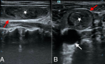
This is a 65-year-old patient who has been on hemodialysis for 5 years with a left humeral-basilic arteriovenous fistula. This fistula is currently in hyper flow estimated at 2300 ml/min associated with three uncomplicated false-aneurysms. There was no thrombosis in the arteries and veins of the upper limb. In order to perform another replacement fistula on the right upper limb, the patient was referred to us for vascular mapping of the right upper limb. This vascular mapping was not favorable to the realization of a native arteriovenous fistula, due to the insufficient caliber of the superficial veins. In addition, exploration of the neck vessels revealed a left jugular thrombosis. The thrombus was hypoechoic oval, measuring 32x8x12mm, floating in the lumen of the internal jugular vein without parietal contact and giving the appearance of a "floating boat" in longitudinal view (A,B). The patient had no other signs of thrombosis and received anticoagulant therapy to prevent the occurrence of another thromboembolic event that could be more deleterious. This case illustrates the importance of scanning the neck vessels during upper extremity vessel ultrasound to look for any abnormalities at this level.
Figure 1: (A) longitudinal view of the jugular vein with the floating thrombus giving the appearance of a floating boat; (B) axial view of the jugulo-carotid vessels showing the floating thrombus without parietal contact. Red arrow: internal jugular vein white arrow: common carotid artery white star: floating thrombus
Search
This article authors
On Pubmed
On Google Scholar
Citation [Download]
Navigate this article
Similar articles in
Key words
Article metrics

