
Post-traumatic pseudoaneurysm of the external carotid artery
Abdel Ilah Drissi Maniani, Najwa Ech-Chrif El Kettani
Corresponding author: Abdel Ilah Drissi Maniani, Neuroradiology Department Head and Neck Hospital of Rabat, Rabat, Morocco 
Received: 09 Jun 2020 - Accepted: 24 Jul 2020 - Published: 18 Mar 2021
Domain: Radiology,Neurology (general)
Keywords: Pseudo aneurysm, external carotid artery, cerebral CT
©Abdel Ilah Drissi Maniani et al. PAMJ Clinical Medicine (ISSN: 2707-2797). This is an Open Access article distributed under the terms of the Creative Commons Attribution International 4.0 License (https://creativecommons.org/licenses/by/4.0/), which permits unrestricted use, distribution, and reproduction in any medium, provided the original work is properly cited.
Cite this article: Abdel Ilah Drissi Maniani et al. Post-traumatic pseudoaneurysm of the external carotid artery. PAMJ Clinical Medicine. 2021;5:68. [doi: 10.11604/pamj-cm.2021.5.68.24184]
Available online at: https://www.clinical-medicine.panafrican-med-journal.com//content/article/5/68/full
Images in clinical medicine 

Post-traumatic pseudoaneurysm of the external carotid artery
Post-traumatic pseudoaneurysm of the external carotid artery
![]() Abdel Ilah Drissi Maniani1,&, Najwa Ech-Chrif El Kettani1
Abdel Ilah Drissi Maniani1,&, Najwa Ech-Chrif El Kettani1
&Corresponding author

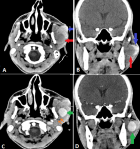
Our case is about a 24-year-old male who consults in an emergency for a left cervical swelling with a history of a penetrating cervical trauma occurred 15 days before. Computed tomography (CT) was performed who shows a hypo dense left cervical collection well limited surmounted by a spontaneously hyper dense hematoma (A, B), with intense enhancement on post contrast at arterial time with individualization of communication with a branch of the external carotid artery (C,D) characteristic of the pseudo aneurysm (PA). The PA or false aneurysm is defined by a rupture of the continuity of the arterial wall with creation of an aneurysmal sac thanks to a pseudo-wall formed by the adjacent structures while maintaining continuity with the nourishing artery, PA carotid arteries are often post-traumatic sometimes other cause are involved (vasculitis, local infection, iatrogenic). The interval between trauma and symptoms is very variable, it results in a pulsating and pulsating cervical swelling on palpation with an audible noise on auscultation, ultrasound shows a cystic mass with a turbulence in the blood flow making the yin-yong sign on doppler examination and to-and-fro waveforms on the pulsed Doppler examination. Computed Tomography generally targets a hypo-dense collection with an intense vascular type enhancement in arterial time and a smooth wall which communicates with a nourishing artery. Magnetic resonance imaging (MRI) allows more characterization (morphology, size of collar, study of collaterality). Conventional angiography is an invasive examination which generally presents a therapeutic interest (stenting, embolization) while the surgical indication is reserved for specific cases.
Figure 1: cerebral CT in axial (A), coronal (B), non contrast section and axial (C), coronal (D), post-contrast section showing a hypo dense left cervical collection well limited (red arrows) surmounted by a spontaneously hyper dense hematoma (blue arrows) with intense enhancement on post contrast at arterial time (green arrows) with individualization of communication with a branch of the external carotid artery (orange arrows)
Search
This article authors
On Pubmed
On Google Scholar
Citation [Download]
Navigate this article
Similar articles in
Key words
Article metrics

