
Chorioretinitis sclopetaria
Karima Madbouhi, Lalla Ouafae Cherkaoui
Corresponding author: Karima Madbouhi, Université Mohammed V Souissi, Ophtalmologie A, Hôpital des Spécialités, Centre Hospitalier Universitaire Rabat, Rabat, Morocco 
Received: 19 Jun 2021 - Accepted: 15 Jul 2021 - Published: 16 Jul 2021
Domain: Ophthalmology
Keywords: Retina, chorioretinitis sclopetaria, hyperpigmentation
©Karima Madbouhi et al. PAMJ Clinical Medicine (ISSN: 2707-2797). This is an Open Access article distributed under the terms of the Creative Commons Attribution International 4.0 License (https://creativecommons.org/licenses/by/4.0/), which permits unrestricted use, distribution, and reproduction in any medium, provided the original work is properly cited.
Cite this article: Karima Madbouhi et al. Chorioretinitis sclopetaria. PAMJ Clinical Medicine. 2021;6:23. [doi: 10.11604/pamj-cm.2021.6.23.30435]
Available online at: https://www.clinical-medicine.panafrican-med-journal.com//content/article/6/23/full
Images in clinical medicine 

Chorioretinitis sclopetaria
Chorioretinitis sclopetaria
![]() Karima Madbouhi1,&,
Karima Madbouhi1,&, ![]() Lalla Ouafae Cherkaoui1
Lalla Ouafae Cherkaoui1
&Corresponding author

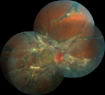
A 26-year-old male who had a history of trauma to the right eye 6 years ago that manifested in damage to the retina, causing chorioretinitis sclopetaria. His visual acuity was counting fingers in his right eye and 20/20 in his left eye. Intraocular pressure was normal in both eyes. Ophthalmic examination, the retina showed a large fibrogliotic lesion associated with hyperpigmentation (figure 1). The left eye and the rest of the physical examination were entirely normal. Sclopetaria is a secondary outcome of a decelerating object passing at a high velocity adjacent to the sclera. While passing close to the globe, after shock forces are generated, rupturing of the choroid and retina occurs (concussion type of injury). The sclera remains intact. Vitreous hemorrhage may occur. The process usually ends up with a white fibrous scar and/or retinal pigmentary alterations that are often the final findings in this situation. The location is mostly at the site adjacent to the trajectory, combined with part of the macula.
Figure 1: image of the posterior pole of the retina showing chorioretinitis sclopetaria: a large fibrogliotic lesion associated with hyperpigmentation
Search
This article authors
On Pubmed
On Google Scholar
Citation [Download]
Navigate this article
Similar articles in
Key words
Tables and figures
Article metrics
PlumX Metrics
Chorioretinitis sclopetaria
