
Imagerie d´un os naviculaire accessoire symptomatique
Amina Alaoui, Nizar El Bouardi
Corresponding author: Amina Alaoui, Radiologue, Département de Radiologie CHP Tata, Tata, Maroc 
Received: 04 Aug 2021 - Accepted: 02 Sep 2021 - Published: 03 Sep 2021
Domain: Radiology,Sport medicine
Keywords: Os naviculaire accessoire, IRM de l’arrière pied, douleurs de l’arrière pied
©Amina Alaoui et al. PAMJ Clinical Medicine (ISSN: 2707-2797). This is an Open Access article distributed under the terms of the Creative Commons Attribution International 4.0 License (https://creativecommons.org/licenses/by/4.0/), which permits unrestricted use, distribution, and reproduction in any medium, provided the original work is properly cited.
Cite this article: Amina Alaoui et al. Imagerie d´un os naviculaire accessoire symptomatique. PAMJ Clinical Medicine. 2021;7:3. [doi: 10.11604/pamj-cm.2021.7.3.31078]
Available online at: https://www.clinical-medicine.panafrican-med-journal.com//content/article/7/3/full
Images in clinical medicine 
Imagerie d´un os naviculaire accessoire symptomatique
Imagerie d´un os naviculaire accessoire symptomatique
Imaging of the symptomatic accessory navicular bone
![]() Amina Alaoui1,&, Nizar El Bouardi2
Amina Alaoui1,&, Nizar El Bouardi2
&Auteur correspondant
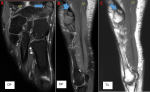
We report through this case a cause that we consider poorly understood and underestimated of foot pain. This is the case of a patient accusing for more than 3 years of mechanical pain in the median surface of the right rear foot, put on multiple orthopedic therapy (insoles) and medication without any improvement. Faced with the non-improvement and the functional repercussions of the pain limiting daily and sporting activity, an magnetic resonance imaging (MRI) of the foot was requested, objectifying the presence of an accessory navicular bone measuring 7.5x5.5 mm, forming a neo-articulation which is the site of arthritic changes associated with edema of the joint edges within the framework of an inflammatory outbreak of osteoarthritis. The final diagnosis of a type II accessory navicular bone forming navicular synchondrosis complicated by congestive osteoarthritis was retained. The treatment consisted of a local infiltration of the synchondrosis by corticosteroids with a good clinical and functional evolution.
Key words: Accessory navicular bone, MRI, pain, rear foot

Nous rapportons à travers ce cas une cause que nous jugeons mal connu et sous-estimée des douleurs du pied. C´est le cas d´un patient accusant depuis plus de 3 ans des douleurs mécaniques de la face médiane de l´arrière pied droit, mise sous multiples moyens thérapeutiques orthopédiques (semelles) et médicamenteuses sans aucune amélioration. Devant la non amélioration et le retentissement fonctionnel de la douleur limitant l´activité quotidienne et sportive, une Imagerie par résonance magnétique (IRM) du pied a été demandé, objectivant la présence d´un os accessoire naviculaire mesurant 7.5x5.5 mm, formant une néo articulation qui est le siège de remaniements arthrosiques associée à un œdème des berges articulaire rentrant dans le cadre d´une poussée inflammatoire d´arthrose. Le diagnostic final d´un os naviculaire accessoire de type II formant une synchondrose naviculaire compliquée d´arthrose en poussée congestive a été retenu. Le traitement a consisté en une infiltration locale de la synchondrose par les corticoïdes avec une bonne évolution clinique et fonctionnelle.
Figure 1: (A,B,C) séquences IRM DP et T1 montrant un os naviculaire accessoire type II (flèche bleu)
Search
This article authors
On Pubmed
On Google Scholar
Citation [Download]
Navigate this article
Similar articles in
Key words
Tables and figures
Article metrics
PlumX Metrics
Imagerie d´un os naviculaire accessoire symptomatique
