Amniotic band disease: a case report
Chawki Mrezguia, Hassine Abouda, Haithem Aloui, Ahmed Halouani, Marwa Souissi, Douaibia Anis, Hadhami Jaouad
Corresponding author: Haithem Aloui, Faculty of Medecine Tunis, Tunis, Tunisia 
Received: 24 Feb 2021 - Accepted: 27 Jan 2022 - Published: 01 Feb 2022
Domain: Obstetrics and gynecology
Keywords: Amniotic band disease, embryo-fetopathies, antenatal diagnosis, prognosis, case report
©Chawki Mrezguia et al. PAMJ Clinical Medicine (ISSN: 2707-2797). This is an Open Access article distributed under the terms of the Creative Commons Attribution International 4.0 License (https://creativecommons.org/licenses/by/4.0/), which permits unrestricted use, distribution, and reproduction in any medium, provided the original work is properly cited.
Cite this article: Chawki Mrezguia et al. Amniotic band disease: a case report. PAMJ Clinical Medicine. 2022;8:25. [doi: 10.11604/pamj-cm.2022.8.25.28547]
Available online at: https://www.clinical-medicine.panafrican-med-journal.com//content/article/8/25/full
Case report 

Amniotic band disease: a case report
Amniotic band disease: a case report
![]() Chawki Mrezguia1, Hassine Abouda1,
Chawki Mrezguia1, Hassine Abouda1, ![]() Haithem Aloui1,&, Ahmed Halouani1, Marwa Souissi1, Douaibia Anis1,
Haithem Aloui1,&, Ahmed Halouani1, Marwa Souissi1, Douaibia Anis1, ![]() Hadhami Jaouad1
Hadhami Jaouad1
&Corresponding author
Amniotic band disease or constriction ring syndrome (CRS) is a variety of birth defects. It is a rare pathology caused by strands of the amniotic sac that separate and entangle the limbs, digits or other parts of the body. Unlike genetic malformations, CRS-related abnormalities are often asymmetrical, polymorphic and do not adhere to any embryological systematization. The complications from the amniotic band disease can range from mild such as amputations of the digits or the limbs, syndactyly and deformities to severe when the band becomes wrapped around the neck, the umbilical cord and the neurological system resulting in fetal death and neural-tube like defects. The strands are often hard to see by ultrasound, and typically the condition is detected indirectly by the constrictions and swellings they cause. Therefore, CRS poses diagnostic difficulties and therapeutic challenges for practitioners. We report two cases of CRS, one of postnatal discovery and one of prenatal discovery. The usefulness is to highlight the diagnostic difficulties of this rare pathology.

Constriction ring syndrome (CRS) is a constellation of fetal morphological abnormalities assosciated with intrauterine amniotic bands. It is a rare pathology [1]. Sentilhes L et al. reports an incidence of between 1/1200 and 1/15,000 live births [2]. Unlike genetic malformations, CRS-related abnormalities are often asymmetrical, polymorphic and do not adhere to any embryological systematization [2]. Although some familial cases have been described, there is no genetic predisposition [3]. Constriction rings of the extremities with resultant auto amputation of digits and craniofacial anomalies are common manifestations of the syndrome. Neural tube like defects are commonly consideded to be secondary to neurulation disorders. Therefore, CRS poses practitioners with difficulties in therapeutic and diagnostic management [4]. We report two cases of CRS, one of which is about a case of a neural tube like defect. The aim of this report is to highlight the challenges associated with the diagnosis and treatement of this rare pathology.

1st case
Patient information: Ms. MH, 36-year-old, with no significant personal pathological history, non-smoking was referred by her gynecologist to our emergency room for childbirth following the spontaneous entry into labor on a term of 39 weeks of amenorrhea and 3 days. It is a spontaneous pregnancy resulting from a non-inbred marriage after a primary hypofertility of 6 years of male origin, conducted without dysgravidia. For his prenatal check-up, the serologies were without abnormalities, it has a first-trimester ultrasound at 11 weeks of amenorrhea and 6 days showing an evolutionary mono-fetal pregnancy consistent with the term nucal with nucal clarity at 1 mm, a morphological ultrasound done at 23 weeks of amenorrhea that is without detectable morphological abnormalities and an ultrasound in the third trimester revealing an evolutionary monofoetal pregnancy with a term-compliant biometric without abnormalities of amniotic fluid or umbilical doppler. The labor was monitored, and the delivery took place on the same day by an emergency caesarean section for pathological detectable foetal heart rhythm.
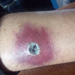
Clinical findings: this is a newborn female. The somatic examination showed a weight of 3400 g, a heart rate of 124 beats/minute, a respiratory rate of 29 cycles/minute and a pulsed oxygen saturation of 96%. At the same time, malformative syndrome was noted in the left hand, with furrows with amputation of the ring and middle finger (Figure 1). In the right hand, there is an index and major syndactyly (Figure 2) and an amputation of the ear. On the left foot, there is a big toe aplasia. Infectious history was negative, with no medication or toxics taken during pregnancy and no similar cases in the family.
Diagnostic assessment: the biological balance showed a normal hemogram and a correct ionogram, a negative C-reactive protein. Thoracoabdominal X-ray was normal with a correct cardiothoracic index. Cardiac and trans-fontanellary ultrasound was without abnormalities. Ultrasound of the abdomen did not object visceral or renal malformations. The diagnosis of constricting ring syndrome was confirmed.
Therapeutic intervention: the newborn was referred for hand surgery.
Follow-up and outcomes: after surgery, the newborn was referred to the pediatric department for better care and support.
2nd case
Patient information: Mrs. A.M, a 26-year-old, a non-inbred marriage, with a family history of a diabetic, primigest mother, referred to our department following the diagnosis of mild preeclampsia at 24 weeks of amenorrhea and whose morphological ultrasound performed at admission objectified an evolutionary monofoetal pregnancy, partial agenesis of the posterior cranial box, presence of the brain in the amniotic fluid without anatomical markers of the structures with evidence of a link between the cranial defect and amniotic bridles. The appearance of the face was difficult to appreciate, with the existence of probable hypertelorism. The rest of the morphology was uncharted. The parents were informed of the pathology of their fetus and its prognosis and a therapeutic termination of the pregnancy was proposed, but the woman refused. The pregnancy maintained until the end of 40 weeks and 1 day, with spontaneous entry into labor and vaginal delivery.
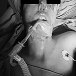
Clinical findings: neonatal examination objectified a newborn male, 3450g of natal weight and several malformations affecting the cephalic extremity (Figure 3): partial absence of brain tissue and cranial arch. The face and eyes are present.
Therapeutic intervention: the newborn was admitted to the neonatology department, he received hydration, parenteral feeding and oxygenation.
Follow-up and outcomes: the trend was marked by death one day after the birth.
Patients perspective
Patient 1: I am happy that doctors all over the world are learning from my child´s case and do not mind my daughter´s condition being discussed. I am thankful for the doctors and the surgeons that took care of me and my child.
Patient 2: I do not want anyone to go through what my husband, I had to. My family is very supportive. I really thank the medical staff for the health care I received and for their effort for taking care of my child. I want to thank them for referring me for a psychiatrist. That helped.
Patients consent: the two patients mentioned in this article gave their consent for the information about their children to be published. They were informed that the Information will be published without their childen´s names attached and every attempt will be made to ensure anonymity. They were aware that the information may be published in a journal which is read worldwide or an online journal. Journals are aimed mainly at health care professionals but may be seen by many non-doctors, including journalists. They were informed that they can withdraw their consent at any time before online publication, but once the information has been committed to publication it will not be possible to withdraw the consent.
The interest of our study is to report rare cases for the clinician. That said, post-partum follow-up is not long enough. This is about the second. Knowing the future of these patients would have provided us with a more comprehensive idea of the pathology. We need to think about the diagnosis of the CRS as early as the first trimester. Abnormalities of the cephalic pole and trunk can be detected by ultrasound at 10-12 WA, while limb abnormalities are diagnosed with a new ultrasound [5]. This pathology is greatly underestimated in its prenatal diagnosis. The prognosis of the CRS depends on the severity of the deformities. In mild forms, surgical or prosthetic palliative treatment can provide an optimal quality of life. On the other hand, severe forms come out of any therapeutic resource, death occurs either in utero or after birth [2]. Amniotic band disease is a relatively rare sporadic condition [6]. This syndrome can be lethal, due to abnormalities that are not compatible with life or by strangulation by an amniotic bridle of the umbilical cord [1]. In the second newborn of our cases, the progression resulted in death. Pathophysiology remains poorly elucidated to this day; multiple different pathological processes have been described [7]. An abnormality of the superficial vascularization of the fetus, in particular the occurrence of hemorrhage, is believed to be the main pathogen [8].
Some anatomical elements (fingers, limbs and skull) of the fetus could externalize, at least partially, from the amniotic cavity, and thus find themselves in close contact with the naked chorion. The outer surface of the amnios (mesodermic) would produce fibrous bands, which would surround and then strangle externalized fetal anatomical elements. These fibrous bands are thus the cause, by a phenomenon of strangulation, of the constriction and amputation lesions found in the CRS [9]. The diagnosis of CRS is possible from the first trimester, and it depends on the nature and severity of the malformations observed [5]. Screening for craniofacial and thoraco-abdominal abnormalities is possible from the first ultrasound, while limb malformations are usually detected during a new ultrasound examination. In our observation, the diagnosis was made only in post-natal for the first case and prenatally during morphological ultrasound for the second case. The CRS is characterized by the asymmetry of abnormalities. Antenatal detection of an amniotic band is not essential to its diagnosis. However, the anatomopathological examination will have to confirm this [10]. Member abnormalities are very varied and are the most common manifestations of the CRS in 65% of cases. Facial malformation is present in 48% of cases, and 77% of cases have at least two abnormalities. Oblique facial clefts are the most described facial abnormalities and are present in 27% of cases [7]. The diagnosis must be evoked before a bundle of arguments namely the constriction or asymmetric amputation of a limb end with lymphedema downstream of constriction, craniofacial asymmetric malformations (encephaloceles, clefts labiopalatins), coelosomies, pseudo-syndactylies and the presence of an amniotic bridle in contact with the injured fetal pole.
Obstetrically, CRS-related malformative abnormalities do not alter the prognosis in relation to the general population [6]. In particular, in cases of craniofacial or visceral malformation, the rate of anomaly of presentation or dystocia is not increased [6]. However, some authors report an increased rate of late miscarriage and prematurity in CRS cases [9]. In the case we reported, caesarean delivery for a cardiofoetal rhythm abnormality for the first case and vaginal delivery for the second, and were conducted without incident, the amount of amniotic fluid was considered normal.
Amniotic disease remains rare, but screening must be routine in the neonatal period. It is an acquired embryofeopathy comprising a set of asymmetric malformations, mainly of interest to the limbs and craniofacial region. Although the pathogenesis of this disease remains controversial, this malformative syndrome requires multidisciplinary management of pregnancy, involving obstetricians, pediatricians, plastic and pediatric surgeons and radiologists. This is essential in order to explain to parent the difficulty of establishing a functional prognosis in antenatal as well as postnatal or even prenatal therapeutic possibilities.
The authors declare no competing interests.
All authors participated in the study. All the authors have read and agreed to the final manuscript.
Figure 1: the left hand showing skin furrows with amniotic amputations of the thumb, index finger and middle finger
Figure 2: image of the right hand showing amniotic amputation of the ear and syndactyly of the index and major
Figure 3: partial absence of brain tissue and cranial arch
- Landolsi A, Jbali S, Helali H, Jablaoui Y, Helali M, Zitouni K et al. Amniotic band syndrome. La Tunisie medicale. 2013;91(5):362-4. PubMed | Google Scholar
- Sentilhes L, Verspyck E, Patrier S, Eurin D, Lechevallier J, Marpeau L. Amniotic band syndrome: pathogenesis, prenatal diagnosis and neonatal management. Journal de Gynecologie, Obstetrique et Biologie de la Reproduction. 2003;32(8 Pt 1):693-704. PubMed | Google Scholar
- de Pablo A, Calb I, Jaimovich L. Congenital constriction bands: amniotic band syndrome. Journal of the American Academy of Dermatology. 1995;32(3):528-9. PubMed | Google Scholar
- Adadi H, Chaara H, Attar I, Jayi S, Alaoui FF, Melhouf MA. Amniotic band syndrome: prenatal diagnosis and management challenges (about 2 cases of lethal malformations). The Pan African Medical Journal. 2019 Mar 13;32:116. PubMed | Google Scholar
- Higginbottom MC, Jones KL, Hall BD, Smith DW. The amniotic band disruption complex: timing of amniotic rupture and variable spectra of consequent defects. The Journal of Pediatrics. 1979;95(4):544-9. PubMed | Google Scholar
- Seeds JW, Cefalo RC, Herbert WN. Amniotic band syndrome. American journal of Obstetrics and Gynecology. 1982;144(3):243-8. PubMed | Google Scholar
- Bouguila J, Ben Khoud N, Ghrissi A, Bellalah Z, Belghith A, Landolsi E et al. Amniotic band syndrome and facial malformations. Revue de Stomatologie et de Chirurgie Maxillo-Faciale. 2007;108(6):526-9. PubMed | Google Scholar
- Daya M. Amniotic band syndrome with persistent sciatic artery: a case report. Annals of Plastic Surgery. 2008;61(5):549-51. PubMed | Google Scholar
- Torpin R. Amniochorionic mesoblastic fibrous strings and amnionic bands: associated constricting fetal malformations or fetal death. American Journal of Obstetrics and Gynecology. 1965;91:65-75. PubMed | Google Scholar
- Lockwood C, Ghidini A, Romero R, Hobbins JC. Amniotic band syndrome: re-evaluation of its pathogenesis. American Journal of Obstetrics and Gynecology. 1989;160(5 Pt 1):1030-3. Google Scholar
Search

This article authors
On Pubmed
On Google Scholar
Citation [Download]
Navigate this article
Similar articles in
Key words
Tables and figures


Article metrics
PlumX Metrics
Amniotic band disease: a case reportRecently from the PAMJ-CM