
Osgood–Schlatter disease: X-ray image
Amina Alaoui, Nizar Bouardi
Corresponding author: Amina Alaoui, Département de Radiologie CHP Tata, Tata, Maroc 
Received: 02 Nov 2021 - Accepted: 06 Jan 2022 - Published: 10 Jan 2022
Domain: Radiology
Keywords: Osgood-Schlatter disease, anterior, knee pain, tibial tubercle
©Amina Alaoui et al. PAMJ Clinical Medicine (ISSN: 2707-2797). This is an Open Access article distributed under the terms of the Creative Commons Attribution International 4.0 License (https://creativecommons.org/licenses/by/4.0/), which permits unrestricted use, distribution, and reproduction in any medium, provided the original work is properly cited.
Cite this article: Amina Alaoui et al. Osgood–Schlatter disease: X-ray image. PAMJ Clinical Medicine. 2022;8:3. [doi: 10.11604/pamj-cm.2022.8.3.32279]
Available online at: https://www.clinical-medicine.panafrican-med-journal.com//content/article/8/3/full
Images in clinical medicine 

Osgood–Schlatter disease: X-ray image
Osgood-Schlatter disease: X-ray image
![]() Amina Alaoui1,&, Nizar Bouardi2
Amina Alaoui1,&, Nizar Bouardi2
&Corresponding author

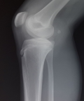
A 14-year-old boy presented a 1-week history of pain in rigth knee. He was active in sports. He reported no specific trauma, fever, or other joint symptoms. A physical examination of the right knee showed mild soft-tissue swelling and tenderness over the tibial tubercle, and the right quadriceps muscle was taut. Plain radiographs of right knee, which were obtained to rule out an avulsion fracture, showed sclerosis and fragmentation of the tibial tubercle. These characteristic findings led to a diagnosis of Osgood-Schlatter disease. Osgood-Schlatter disease (OSD) is one of the most common causes for anterior knee pain in children and adolescents resulting from a traction apophysitis of the tibial tubercle. While a peak in boys aged 12-15 years old was well documented, there seems to be no difference in sex distribution nowadays. This may result from increased participation of young females in high-impact sports. OSD is a mostly self-limiting apophysitis of the tibial tubercle in young active patients with open physis.
Figure 1: lateral view radiograph showing acute signs of unilateral Osgood-Schlatter disease: mild swelling as well as fragmentation of the anterior aspect of the tibial tubercle
Search

This article authors
On Pubmed
On Google Scholar
Citation [Download]
Navigate this article
Similar articles in
Key words
Article metrics
PlumX Metrics
Osgood–Schlatter disease: X-ray imageRecently from the PAMJ-CM


