
A conservative approach towards idiopathic chondrolysis of hip: a case report
Sanika Pravin Gade, Om Chandrakant Wadhokar, Pratik Arun Phansopkar
Corresponding author: Pratik Arun Phansopkar, Department of Musculoskeletal Physiotherapy, Ravi Nair Physiotherapy College, DattaMeghe Institute of Medical Sciences, Sawangi(M), Wardha, Maharashtra, India 
Received: 12 Feb 2022 - Accepted: 11 Mar 2022 - Published: 17 Mar 2022
Domain: Physical medicine and rehabilitation or Physiatry
Keywords: Idiopathic chondrolysis of hip, avascular necrosis, physical therapy, rehabilitation, case Report
©Sanika Pravin Gade et al. PAMJ Clinical Medicine (ISSN: 2707-2797). This is an Open Access article distributed under the terms of the Creative Commons Attribution International 4.0 License (https://creativecommons.org/licenses/by/4.0/), which permits unrestricted use, distribution, and reproduction in any medium, provided the original work is properly cited.
Cite this article: Sanika Pravin Gade et al. A conservative approach towards idiopathic chondrolysis of hip: a case report. PAMJ Clinical Medicine. 2022;8:45. [doi: 10.11604/pamj-cm.2022.8.45.33744]
Available online at: https://www.clinical-medicine.panafrican-med-journal.com//content/article/8/45/full
Case report 

A conservative approach towards idiopathic chondrolysis of hip: a case report
A conservative approach towards idiopathic chondrolysis of hip: a case report
![]() Sanika Pravin Gade1,&,
Sanika Pravin Gade1,&, ![]() Om Chandrakant Wadhokar1,
Om Chandrakant Wadhokar1, ![]() Pratik Arun Phansopkar1
Pratik Arun Phansopkar1
&Corresponding author
Chondrolysis of the hip causes pain and a limping as articular cartilage space is gradually lost due to an unknown but suspected inflammatory process. It could be idiopathic or due to another hip condition, such as slipped capital femoral epiphysis. It is a well-known complication of a slipped capital femoral epiphysis, although it can also occur as a result of trauma, septic arthritis, immobilization, or juvenile idiopathic arthritis. Idiopathic chondrolysis of the hip is a disorder that causes the articular cartilage of the hip joint to degrade spontaneously. The most common clinical manifestation is pain at the affected hip joint, which is linked to increasing joint stiffness and a significant loss of range of motion and function. Idiopathic chondrolysis of hip is a rare condition with Incidence around 8.2% percent. In this case report we are presenting a 12-year-old boy came with complaint of pain, stiffness and restricted range of movement at left hip joint followed by trauma. On the basis of physical and investigatory findings, the patient was diagnosed as a case of idiopathic chondrolysis of hip associated with avascular necrosis grade 4 according to Ficat and Arlet classification. Radiological findings were evident of reduced joint space of left hip and slipped capital epiphysis, this is known to be the hallmark finding to confirm the diagnosis of idiopathic chondrolysis of hip. The patient was conservatively managed with non-steroidal anti-inflammatory medicines, vigorous physiotherapy. Chrondolysis is usually managed with bed rest, traction and non-steroidal anti-inflammatory drugs, hence the aim of this case report was to incorporate an elaborate and efficiency of physical Therapy along with medical management to promote the recovery, aid early return to activities of daily living and improve quality of life.

Chondrolysis is a condition in which the hyaline cartilage of the acetabulum and femoral head begins to deteriorate progressively, which may be idiopathic or, more commonly, secondary to other pathological processes [1]. Idiopathic chondrolysis of the hip is a rare disorder with unidentified causes which is manifested by pain, stiffness and an antalgic pattern of gait [2]. The necrosis is usually associated with slipped capital femoral epiphysis, trauma or sepsis. Concentric narrowing of joint space is the hallmark radiographic finding of this condition [3]. Incidence of chondrolysis in slipped capital femoral epiphysis is 8.2%. It is most common in Asian or African-American female teenagers, and it is frequently mono-articular (60% affecting right side). In 5% of patients, there is a bilateral chondrolysis of hip [4]. To make a differential diagnosis, medical imaging techniques are required, and biological markers for inflammation and infection should be assessed [5]. This report presents a rare case of a 12-year-old boy whose symptoms, physical examination findings and investigatory results were directed towards idiopathic hip chondrolysis associated with grade 4 avascular necrosisaccording to Ficat and Arlet classification. The aim of this report is to highlight the importance of physical therapy in the management of the disorder in order to facilitate early ambulation.

Patient information: patient in the present case is a 12-year-old boy, resident of Chandrapur, reported to the orthopedic department with complaints of pain andrestriction of movements of left hipwhich was followed by trauma and abrupt jerk to left hip while riding a bicycle 9 months back. Immediately after injury, the patient experienced pain and swelling over the left hip joint, along with difficulty in movement. Diffuse pain was present over the left hip, the pain was sudden in onset and associated with swelling. Pain was dull aching in nature and continuous throughout the day. The intensity of pain was rated as 8/10 on numerical pain rating scale as per the patient. Pain used to get aggravated on movement and relieved on rest and medications. Patient has been walking with a limping over the left side. Patient then went to a private clinic where he was managed conservatively with medications as the pain was persistent he was referred to AVBRH hospital where investigations like X-ray and Magnetic Resonance Imaging (MRI) were done. On X-ray, reduced joint space was evident of left hip and MRI findings were suggestive of avascular necrosis of left femoral head due to slipped epiphysis stage 4. These findings along with clinical manifestations confirmed the diagnosis of idiopathic chondrolysis of hip.
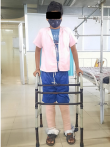
Clinical findings: the patient was assessed in supine lying with pulse rate 85 beats/min, respiratory rate 21 breaths/minute, blood pressure was 122/80 mmHg. The attitude of left lower limb was 20 degrees of abduction slight external rotation knee completely extended and ankle slightly planter flexed. Skin traction was applied bilaterally on leg. On inspection swelling absent, the patient´s right ASIS was higher than compared to the left ASIS, posture was assessed in standing position which revealed increased lumbar lordosis and functional scoliosis (Figure 1) the patient walks with limping on the left side. On palpation, grade 2 tenderness is present at the greater tubercle of the left hip. On girth assessment, there was 5 cm difference in girth at the mid-thigh level. On limb length assessment, the patient presented with 4 cm difference in apparent limb length. All higher functions were intact. On Examination the Range of motion of the right side was full and functional the ROM of left lower limb is mentioned in the (Table 1). Manual muscle testing of right lower limb was grade 5 for all musculature, the strength of left lower limb is mentioned in (Table 2).
Timeline of current episode: April 11, 2021: date of injury/ incidence, followed by patient visit to private clinic, ultrasonography was performed evident of semimembranosus and semitendinosus tear. July 2021: patient on ayurvedic treatment for 2 months. January 16, 2022: patient admitted to orthopedic ward. January 17, 2022: X-ray was performed, suggestive of reduced joint space of left hip. January 22, 2022: conservative management with balanced below knee traction. January 25, 2022: physiotherapy rehabilitation commenced.
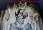
Diagnostic assessment: an AP X-ray on both hip was taken which revealed significant reduction in the left hip joint space, along with right iliac at higher level as compared to the left hip with slightly slipped left femoral epiphysis (Figure 2). Magnetic resonance imaging (MRI) bilateral hip with pelvis was also performed; evidence of; the left femoral physeal plate is narrow with decreased height of femoral head; evidence of left protrusion acetabula; minimal left side hip joint effusion; impression of MRI revealed avascular necrosis of left femoral due to slipped epiphysis stage 4.
Diagnosis: evaluations of all the findings are in confirmatory with idiopathic hip chondrolysis. The left femoral head was detected with stage 4 avascular necrosis.
Therapeutic interventions: a conservative approach was started for management of the patient; the treatment included non-steroidal anti-inflammatory drugs along with bilateral skin traction and a well-structured physical therapy protocol was initiated three days after the application of skin traction. The patient was educated about the condition and its prognosis, patient and care taker were advised to avoid high impact activities such as jumping and running on hard surface joint protection techniques were taught to the patient and the care taker, further physical therapy management is mentioned in (Table 3).
Follow up and outcome of interventions: outcome measures used for this case were goniometry for range of motion (Table 4), manual muscle testing to assess the strength of the muscles (Table 5) and numerical pain rating scale to assess pain; preoperatively, patient rated pain as 8/10, and post-operatively he rated pain as 3/10. All these values were taken before the initiation of physiotherapy regimen and compared with values taken at the end of protocol.
Patient perspective: I had an injury to my hip 9 months back for which I have taken various medications, but the pain was not improved after admission to AVBRH hospital and receiving medical therapy and vigorous physical therapy have helped me by alleviate pain and I gained back the confidence in daily activities.
Informed consent: the information regarding the case report was given to the patient and an informed oral consent was obtained from the patient.
Chondrolysis, meaning destruction of cartilage, of the hip joint could be idiopathic, or secondary to other pathological processes. Secondary causes in children may be the consequence of a wide variety of conditions including slipped capital femoral epiphysis, trauma, infection or juvenile idiopathic arthritis [6]. Idiopathic chondrolysis of the hip is a condition that affects adolescents and is marked by the slow degeneration of the hyaline cartilage that surrounds the head of the femur. It eventually reduces hip joint space and, as a result, causes pain, restricts hip joint range of motion [7]. The condition is extremely rare and mostly affects adolescents. Conservative therapies, such as pain medication, traction and physical therapy, and the use of crutches to minimize weight-bearing, are preferred when an adolescent is diagnosed with the condition [8]. The patient reported in this case is an adolescent boy who is diagnosed with idiopathic chondrolysis of hip, which is supposed to be secondary to trauma followed by avascular necrosis of head of femur of left hip. The patient complained of pain at the hip joint, difficulty in ambulation, and restricted range of movements at the affected hip. Investigations like X-ray and MRI confirmed the diagnosis of the condition. X-ray findings were evident of narrowed joint space and slipping of femoral head, which is known to be the trademark finding. MRI findings showed evidence of the left femoral physical plate narrowed with decreased height of femoral head and evidence of left protrusionacetabuli, idiopathic chondrolysis of the hip should be detected in patients who have a gradual loss of hip joint cavity and protrusion acetabuli at the same time [9]. Management of this disorder consists of non-steroidal anti-inflammatory drug, bed rest and skin traction in order to limit workload on the hip joint, and use of crutches while walking [10]. During the first several months, physical therapy is important to ensure the joint range of motion [11]. Fixation of the hip joint should be avoided since it can lead to joint stiffness and fibrous ankylosis [12]. A total hip arthroplasty may also be useful in managing pain in individuals under the age of 20, but it has been observed that it may also produce durability issues, so it should be carefully considered.
We conclude that a multidisciplinary approach involving medical therapy and well-designed physical therapy regimen showed a significant improvement in the patient condition by reducing pain, and improving functional mobility of the patient diagnosed with grade 4 avascular necrosis followed by idiopathic chondrolysis.
The authors declare no competing interests.
All authors contributed equally in drafting the manuscript.
Table 1: pre-rehabilitation range of motion
Table 2: pre-rehabilitation manual muscle testing
Table 3: physiotherapy treatment protocol
Table 4: pre and post rehabilitation range of motion
Table 5: pre and post rehabilitation muscle strength
Figure 1: AP view of patient
Figure 2: AP view X-ray of bilateral hip joint
- Rachinsky I, Boguslavsky L, Cohen E, Hertzanu Y, Lantsberg S. Bilateral idiopathic chondrolysis of the hip: a case report. Clinical Nuclear Medicine. 2000;25(12). PubMed | Google Scholar
- Segaren N, Abdul-Jabar HB, Segaren N, Hashemi-Nejad A. Idiopathic chondrolysis of the hip: presentation, natural history and treatment options. Journal of Pediatric Orthopedics Part B. 2014;23(2). PubMed | Google Scholar
- Dechosilpa C, Mulpruek P, Woratanarat P, Thiabratana P. Idiopathic chondrolysis of the hip (ICH): report of three cases. Malays Orthop J. 2014;8(3):30-32. PubMed | Google Scholar
- Pioger C, Saithna A, Kandhari V, Thaunat M, Vieira TD, Freychet B et al. Risk factors for rapid chondrolysis after partial lateral meniscectomy: a scoping review of the literature. Orthop J Sports Med. 2021;9(2):2325967120981777. PubMed | Google Scholar
- Dendane M-A, Amrani A, Abouqal R, Gourinda H, Ahid S. Factors influencing the development of chondrolysis in children treated for slipped capital femoral epiphysis. Arch Pediatr. 2014;21(8):821-826. PubMed | Google Scholar
- Joseph B, Pydisetty RKV. Chondrolysis and the stiff hip in Perthes´ Disease: an immunological study. Journal of Pediatric Orthopaedics. 1996;16(1):15-19. PubMed | Google Scholar
- Johnson K, Haigh SF, Ehtisham S, Ryder C, Gardner-Medwin J. Childhood idiopathic chondrolysis of the hip: MRI features. Ped Radiol. 2003;33(3):194-199. PubMed | Google Scholar
- Thacker MM, Feldman DS, Madan SS, Straight JJ, Scher DM. Hinged distraction of the adolescent arthritic hip. Journal of Pediatric Orthopaedics. 2005;25(2):178-182. PubMed | Google Scholar
- Appleyard DV, Schiller JR, Eberson CP, Ehrlich MG. Idiopathic chondrolysis treated with etanercept. Orthopedics. 2009;32(3):214. PubMed | Google Scholar
- Amarnath C, Muthaiyan P, Mary TH, Mohanan S, Gopinathan K. Idiopathic chondrolysis of hip in children: new proposal and implication for radiological staging. Indian J Radiol Imaging. 2018;28(2):205-213. PubMed | Google Scholar
- Chechik O, Dekel S. Bilateral chondrolysis of the hip following liver transplantation. Skeletal Radiol. 2009;38(3):297-300. PubMed | Google Scholar
- Laor T, Crawford AH. Idiopathic chondrolysis of the hip in children: early MRI findings. AJR Am J Roentgenol. 2009;192(2):526-531. PubMed | Google Scholar
Search
This article authors
On Pubmed
On Google Scholar
Citation [Download]
Navigate this article
Similar articles in
Key words
Tables and figures
Article metrics

