Carcinoma en-cuirasse a rare cutaneous presentation of breast malignancy
Vadlamudi Nagendra, Harshith Gowda
Corresponding author: Harshith Gowda, Department of Radio-Diagnosis, Jawaharlal Nehru Medical College, Sawangi (Meghe), Wardha, Maharashtra, India 
Received: 28 Jun 2022 - Accepted: 04 Jul 2022 - Published: 05 Jul 2022
Domain: Radiology,Dermatology,Oncology
Keywords: Neurological, complications, ankylosing, spondylitis
©Vadlamudi Nagendra et al. PAMJ Clinical Medicine (ISSN: 2707-2797). This is an Open Access article distributed under the terms of the Creative Commons Attribution International 4.0 License (https://creativecommons.org/licenses/by/4.0/), which permits unrestricted use, distribution, and reproduction in any medium, provided the original work is properly cited.
Cite this article: Vadlamudi Nagendra et al. Carcinoma en-cuirasse a rare cutaneous presentation of breast malignancy. PAMJ Clinical Medicine. 2022;9:21. [doi: 10.11604/pamj-cm.2022.9.21.36115]
Available online at: https://www.clinical-medicine.panafrican-med-journal.com//content/article/9/21/full
Images in clinical medicine 
Carcinoma en-cuirasse a rare cutaneous presentation of breast malignancy
Carcinoma en-cuirasse a rare cutaneous presentation of breast malignancy
![]() Vadlamudi Nagendra1,
Vadlamudi Nagendra1, ![]() Harshith Gowda1,&
Harshith Gowda1,&
&Corresponding author

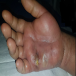
A 65-year-old female complained of a lump in the right breast for 6 months, gradually increasing from the size of pea to the size of a lemon. The lesion was involving and causing retraction of the nipple areola complex, with ulceration and hemorrhagic discharge. On contrast computed tomography (CT) of the thorax, there was an ill-defined heterogeneously enhancing mass lesion with central hypodense area and speculated margins in the retro-areolar region, measuring approximately 7 x 3.2 x 1.9 cm. The lesion was causing architectural distortion of nipple areolar complex with cutaneous and subcutaneous extensive thickening and fat stranding. Computed tomography of the abdomen also revealed multiple liver metastasis. Biopsy from right breast lump revealed invasive ductal carcinoma. Patient was advised palliative chemotherapy. Carcinoma en-cuirasse is a rare presentation of either scirrhous breast carcinoma or cutaneous metastasis which usually spreads through lymphatics. It is highly invasive slow growing variant showing extensive fibrous and dense connective tissue encasing the chest wall. Stromal reaction has been interpreted as either promoting tumor angiogenesis and physical fibrillar matrix components or creating a barrier to halt tumor spread. The dense stromal matrix and less vascularity may be the reason why the tumor is relatively resistant to systemic chemotherapy.
Figure 1: A) large nodular breast mass involving nipple areolar complex. It is associated with serosanguinous and hemorrhagic discharging ulcers; B) contrast CT thorax - an ill-defined heterogeneously enhancing mass lesion with central hypodense area and speculated margins in the retro-areolar region with cutaneous and subcutaneous extensive thickening and fat stranding
Search

This article authors
On Pubmed
On Google Scholar
Citation [Download]
Navigate this article
Similar articles in
Key words

Article metrics
Recently from the PAMJ-CM