
A case of lunatomalacia
Parth Shah
Corresponding author: Parth Shah, Department of Orthopedics, Datta Meghe Institute of Medical Sciences, Wardha, Maharashtra, India 
Received: 08 Jul 2022 - Accepted: 13 Aug 2022 - Published: 17 Aug 2022
Domain: Orthopedic surgery
Keywords: Lunatomalacia, Kienbock's disease, avascular necrosis
©Parth Shah et al. PAMJ Clinical Medicine (ISSN: 2707-2797). This is an Open Access article distributed under the terms of the Creative Commons Attribution International 4.0 License (https://creativecommons.org/licenses/by/4.0/), which permits unrestricted use, distribution, and reproduction in any medium, provided the original work is properly cited.
Cite this article: Parth Shah et al. A case of lunatomalacia. PAMJ Clinical Medicine. 2022;9:38. [doi: 10.11604/pamj-cm.2022.9.38.36262]
Available online at: https://www.clinical-medicine.panafrican-med-journal.com//content/article/9/38/full
Images in clinical medicine 

A case of lunatomalacia
A case of lunatomalacia
![]() Parth Shah1,&
Parth Shah1,&
&Corresponding author

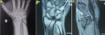
Lunatomalacia is a condition characterized by avascular necrosis of the lunate bone. It is also known as osteonecrosis, Kienbock's disease, and aseptic or ischemic necrosis of the lunate. Although the mechanisms by which this disorder develops are not fully understood, compromise of the bone vasculature is the most commonly proposed cause. Both extraosseous and intraosseous blood vessels supply the lunate bone. Vessels that enter the lunate through the dorsal and volar poles of the bone make up the extraosseous blood supply. Pathomechanics where acute trauma or repetitive minor trauma leads to direct vessel break and ligament disruption causes blood supply interruption and bone necrosis. A 31-year-old male presented to the orthopedics outpatient department with pain and swelling over the right wrist since 1 year. Patient gave alleged history of trauma to wrist while lifting heavy object and Pain was sudden in onset, gradually progressive in nature and dull aching type which got aggravated by lifting weights and got relieved by taking rest and medication. On local examination, tenderness presented over radial styloid, ulna styloid and medial radio-ulnar joint. Wrist range of motion (ROM) range normal and terminally painful. Fovea sign - positive, suggest triangular fibrocartilage complex injury. Pain on ulnar deviation of wrist. The patient was managed with a form of splinting.
Figure 1: (A) X-ray of right wrist shows sclerosis of lunate and negative ulnar variance; (B) coronal T1-weighted mitral regurgitation (MR) image showed diffuse hypo intensity of lunate bone; (C) MR image showed coronal fracture of lunate bone
Search
This article authors
On Pubmed
On Google Scholar
Citation [Download]
Navigate this article
Similar articles in
Key words
Article metrics
PlumX Metrics
A case of lunatomalacia
