Fibrothécome ovarien ou léiomyosarcome: l´extemporané tranche
Taamallah Meriem, Salah Houda
Corresponding author: Taamallah Meriem, Department of Gynecology and Obstetrics, Mohamed Tlatli Hospital, Faculty of Medicine of Tunis, University of Tunis El Manar, Tunis, Tunisia 
Received: 22 Jun 2026 - Accepted: 16 Aug 2026 - Published: 26 Oct 2026
Domain: Gynecology
Keywords: Fibrothécome ovarien, examen extemporané, système O‑RADS, masse annexielle, léiomyosarcome
Funding: Funding statement file not available - Manually update the funding statement
©Taamallah Meriem et al. PAMJ Clinical Medicine (ISSN: 2707-2797). This is an Open Access article distributed under the terms of the Creative Commons Attribution International 4.0 License (https://creativecommons.org/licenses/by/4.0/), which permits unrestricted use, distribution, and reproduction in any medium, provided the original work is properly cited.
Cite this article: Taamallah Meriem et al. Fibrothécome ovarien ou léiomyosarcome: l´extemporané tranche. PAMJ Clinical Medicine. 2026;22:13. [doi: 10.11604/pamj-cm.2026.22.13.54137]
Available online at: https://www.clinical-medicine.panafrican-med-journal.com//content/article/22/13/full
 PDF in process
Images in clinical medicine 

Fibrothécome ovarien ou léiomyosarcome: l´extemporané tranche
Fibrothécome ovarien ou léiomyosarcome: l'extemporané tranche
Ovarian fibrothecoma mimicking leiomyosarcoma: decisive role of frozen section
 Taamallah Meriem1,&, Salah Houda1
&Auteur correspondant
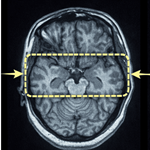
We report the case of a 55-year-old premenopausal woman, G2P2, who presented with perimenopausal menometrorrhagia. Pelvic ultrasound followed by magnetic resonance imaging (MRI) revealed a uterus with multiple leiomyomas, including three distinct lesions: a 4-cm fundal FIGO type 2-5 leiomyoma, a 5-cm anterior isthmic FIGO type 6 leiomyoma, and a 4-cm posterior FIGO type 6 leiomyoma with disruption of the serosa. A 7-cm right parauterine mass with solid and cystic components, classified as O-RADS 4, was also identified. In view of the clinical and radiological findings, leiomyosarcoma could not be ruled out. An initial laparoscopic approach with planned right adnexectomy and intraoperative frozen section examination was decided upon. Intraoperative histopathological examination unexpectedly indicated an ovarian fibrothecoma, a benign ovarian stromal tumor, in the presence of moderate ascites and without peritoneal carcinomatosis. Consequently, the surgical strategy was adapted intraoperatively, changing from extensive oncologic resection to total hysterectomy with bilateral salpingo oophorectomy, which also provided definitive treatment for the menometrorrhagia. Gross examination of the right adnexectomy specimen revealed a firm, yellowish mass with a whorled appearance and an intact capsule. Final histopathological examination confirmed a benign ovarian fibrothecoma and uterine leiomyomas without atypia or evidence of malignancy. This case illustrates the crucial role of frozen section examination in the surgical management of O-RADS 4 adnexal masses in perimenopausal women with concomitant uterine disease. It allowed appropriate therapeutic de-escalation, avoiding unnecessary extensive oncologic surgery while ensuring complete resolution of the symptoms.
Keywords: Ovarian fibrothecoma, frozen section, O-RADS system, adnexal mass, leiomyosarcoma

Nous rapportons le cas d'une patiente âgée de 55 ans, non ménopausée, G2 P2, qui a consulté pour des ménométrorragies péri-ménopausiques; une échographie puis une IRM pelvienne ont objectivé un utérus polymyomateux comportant trois léiomyomes distincts - un fibrome fundique de 4 cm de type 2/5, un fibrome antéro-isthmique de 5 cm de type 6 et un fibrome postérieur de 4 cm de type 6 avec effraction de la séreuse - associé à une masse latéro-utérine droite de 7 cm à composante solido-kystique classée O-RADS 4, pour laquelle le diagnostic de léiomyosarcome ne pouvait être éliminé devant le contexte. Une approche première par cœlioscopie avec annexectomie droite programmée et examen extemporané a été décidée; l'analyse peropératoire a conclu de manière inattendue à un fibrothécome ovarien, tumeur bénigne du stroma ovarien avec une ascite de moyenne abondance, sans carcinose péritonéale. En conséquence, la stratégie chirurgicale a été adaptée en temps réel, passant d'une résection oncologique élargie à une hystérectomie totale avec annexectomie bilatérale définitive, permettant également de traiter radicalement les ménométrorragies. L'examen macroscopique de la pièce d'annexectomie droite montrait une masse ferme, jaunâtre, à contours tourbillonnants et à capsule intacte, et l'histologie définitive a confirmé le fibrothécome bénin ainsi que des fibromes utérins sans atypies ni signes de malignité. Ce cas illustre parfaitement l'apport crucial de l'extemporané dans la prise en charge chirurgicale des masses annexielles O-RADS 4 chez la patiente périménopausée porteuse d'une pathologie utérine associée, en permettant une désescalade thérapeutique adaptée, évitant une chirurgie carcinologique inutile tout en assurant une résolution complète des symptômes.
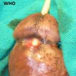
Figure 1: laparoscopique peropératoire
Search

This article authors
On Pubmed
On Google Scholar
Citation [Download]
Navigate this article
Similar articles in
Key words
Tables and figures

Article metrics
Recently from the PAMJ-CM