Respiratory epithelial adenomatoid hamartoma of the nasopharynx: a case report
Oumayma Leassal, Cedric Ango, Mounir Hmidi, Hicham Attifi
Corresponding author: Oumayma Leassal, ENT and Head and Neck Surgery Department, Military Hospital Moulay Ismail, Meknes, Morocco 
Received: 29 Mar 2026 - Accepted: 13 Oct 2026 - Published: 05 Nov 2026
Domain: Otolaryngology (ENT)
Keywords: Respiratory epithelial adenomatoid hamartoma, nasopharynx, differential diagnosis, endoscopic management, case report
Funding: This work received no specific grant from any funding agency in the public, commercial, or non-profit sectors.
©Oumayma Leassal et al. PAMJ Clinical Medicine (ISSN: 2707-2797). This is an Open Access article distributed under the terms of the Creative Commons Attribution International 4.0 License (https://creativecommons.org/licenses/by/4.0/), which permits unrestricted use, distribution, and reproduction in any medium, provided the original work is properly cited.
Cite this article: Oumayma Leassal et al. Respiratory epithelial adenomatoid hamartoma of the nasopharynx: a case report. PAMJ Clinical Medicine. 2026;22:17. [doi: 10.11604/pamj-cm.2026.22.17.52433]
Available online at: https://www.clinical-medicine.panafrican-med-journal.com//content/article/22/17/full
 PDF in process
Case report 

Respiratory epithelial adenomatoid hamartoma of the nasopharynx: a case report
Respiratory epithelial adenomatoid hamartoma of the nasopharynx: a case report
 Oumayma Leassal1,&, Cedric Ango1, Mounir Hmidi1, Hicham Attifi1
&Corresponding author
Respiratory epithelial adenomatoid hamartoma (REAH) is a rare benign lesion of the sinonasal tract, most commonly arising from the posterior nasal septum and frequently associated with chronic inflammatory conditions. Nasopharyngeal localization is exceptional and may mimic other benign or malignant nasopharyngeal masses, leading to diagnostic challenges. We report the case of a 45-year-old man presenting with progressive bilateral nasal obstruction and hyponasal speech evolving over eight months. Nasal endoscopy revealed a smooth polypoid mass arising from the right posterolateral wall of the nasopharynx, obstructing the ipsilateral choana and extending toward the oropharynx. Computed tomography showed a well-defined polylobulated soft tissue lesion without bone destruction or invasion of adjacent structures. Endoscopic biopsy suggested an adenomatoid epithelial hamartoma. Complete surgical excision was performed using a combined endoscopic endonasal and transoral approach. Histopathological examination confirmed the diagnosis of REAH. The postoperative course was uneventful, with no recurrence after 12 months of follow-up. This case highlights the importance of considering REAH in the differential diagnosis of nasopharyngeal masses to avoid misdiagnosis and unnecessary aggressive treatment.

Respiratory epithelial adenomatoid hamartoma (REAH) is a rare benign lesion of the sinonasal tract characterized by glandular proliferation lined by pseudostratified ciliated respiratory epithelium. Wenig and Heffner first described it as a distinct clinicopathological entity in 1995 [1]. Respiratory epithelial adenomatoid hamartoma (REAH) most frequently arises from the posterior nasal septum and is often associated with chronic inflammatory sinonasal conditions, particularly chronic rhinosinusitis and nasal polyposis [2,3]. Nasopharyngeal localization of REAH remains exceptional [4]. Because of its unusual location and tumor-like appearance, it may mimic other benign or malignant nasopharyngeal masses, making diagnosis challenging and exposing patients to the risk of overtreatment [3,4]. We report a rare case of nasopharyngeal REAH presenting as an extensive nasopharyngeal mass and successfully managed by combined endoscopic endonasal and transoral surgical excision.

Patient information: a 45-year-old male with no significant past medical or surgical history presented to our department with progressive bilateral nasal obstruction, more pronounced on the right side, associated with hyponasal speech evolving over approximately eight months. The patient denied epistaxis, purulent rhinorrhea, facial pain, anosmia, headache, or weight loss. There was no history of allergic rhinitis, chronic rhinosinusitis, or previous nasal surgery, and no known exposure to environmental or occupational risk factors.
Clinical findings: on general examination, the patient was in good overall condition with no signs of systemic illness. Nasal endoscopy revealed a well-circumscribed, polypoid mass with a smooth, regular surface arising from the right posterolateral wall of the nasopharynx. The lesion was non-ulcerated, non-bleeding on contact, and obstructed the ipsilateral choana (Figure 1). It extended inferiorly behind the soft palate and was visible on oropharyngeal examination (Figure 2). There was no evidence of mucosal infiltration or suspicious features suggestive of malignancy. No cervical lymphadenopathy was noted. The remainder of the otorhinolaryngological examination was unremarkable.
Timeline of current episode: the timeline of this case is summarized as follows: month 0: Onset of nasal obstruction and hyponasal speech. Month 6: progressive worsening of symptoms. Month 8: ENT consultation and nasal endoscopy. Month 8: computed tomography (CT) scan performed. Month 9: endoscopic biopsy. Month 10: surgical excision. Month 22: follow-up at 12 months with no recurrence.
Diagnostic assessment: computed tomography (CT) of the paranasal sinuses showed a well-defined, homogeneous, polylobulated soft tissue mass occupying the nasopharynx, lateralized to the right, and protruding into the ipsilateral choana (Figure 3). The lesion exhibited no bone erosion, calcifications, or invasion of adjacent anatomical spaces, including the parapharyngeal and retropharyngeal spaces. Based on clinical and radiological findings, the initial differential diagnosis included antrochoanal polyp, inverted papilloma, Thornwaldt cyst, and nasopharyngeal carcinoma. An endoscopic biopsy was performed to establish a definitive diagnosis. Histopathological examination suggested an adenomatoid epithelial hamartoma.
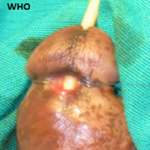
Therapeutic intervention: the patient underwent complete surgical excision under general anesthesia using a combined endoscopic endonasal and transoral approach. This approach was chosen due to the size of the lesion and its inferior extension, allowing optimal exposure and safe en bloc resection. The excised specimen measured approximately 5.5 cm in its greatest dimension (Figure 4).
Follow-up and outcomes: definitive histopathological examination confirmed the diagnosis of respiratory epithelial adenomatoid hamartoma (REAH). Microscopic analysis revealed glandular proliferation lined by pseudostratified ciliated respiratory epithelium embedded in an edematous stroma, without cytologic atypia or mitotic activity. The postoperative course was uneventful. No immediate or delayed complications were observed. At 12 months of follow-up, the patient remained asymptomatic, with complete resolution of nasal obstruction and hyponasal speech. Endoscopic examination revealed no evidence of local recurrence.
Patient perspective: the patient reported a marked improvement in nasal breathing and daily comfort after surgery, with significant enhancement in quality of life, and expressed satisfaction with the management and outcome.
Informed consent: written informed consent was obtained from the patient for publication of this case report and the accompanying images.
Respiratory epithelial adenomatoid hamartoma (REAH) is a rare benign lesion of the sinonasal tract characterized by glandular proliferation lined by pseudostratified ciliated respiratory epithelium. Wenig et al. first described it as a distinct clinicopathological entity in 1995 in a series of 31 cases involving the sinonasal tract and nasopharynx [1]. Respiratory epithelial adenomatoid hamartoma (REAH) is considered an uncommon lesion that typically affects adults in the fifth and sixth decades of life, with a slight male predominance [2,3]. It most frequently arises from the posterior nasal septum and is commonly associated with chronic inflammatory sinonasal diseases, particularly chronic rhinosinusitis and nasal polyposis [2,4]. In contrast, primary localization within the nasopharynx remains exceptionally rare, with only a limited number of cases reported in the literature [4,5-8]. The pathogenesis of REAH remains incompletely understood. It is generally considered a hamartomatous proliferation rather than a true neoplasm, likely related to chronic inflammatory stimulation of the sinonasal mucosa [2]. Chronic inflammation may induce epithelial invagination and glandular proliferation, resulting in the characteristic adenomatoid architecture observed histologically. This hypothesis is supported by the frequent association of REAH with chronic rhinosinusitis and nasal polyposis reported in the literature [2,4].
Clinically, REAH usually presents with nonspecific symptoms, most commonly nasal obstruction, which may be associated with rhinorrhea, hyposmia, or headache [2,3]. In cases involving the nasopharynx, symptoms are mainly related to airway obstruction and may include nasal blockage, snoring, or hyponasal speech. In our case, the patient presented with progressive nasal obstruction and hyponasal speech, consistent with previously reported observations. Radiological imaging plays an important role in the evaluation of sinonasal masses. Computed tomography typically demonstrates a well-defined soft tissue lesion without bone destruction or aggressive features, which may help differentiate REAH from malignant tumors [3]. In the present case, CT findings showed a well-circumscribed polylobulated mass without bone erosion or invasion of adjacent structures, supporting a benign process.
The differential diagnosis of a nasopharyngeal mass includes both benign and malignant entities, such as antrochoanal polyp, Thornwaldt cyst, inverted papilloma, and nasopharyngeal carcinoma [3,4]. Although clinical and radiological features may provide useful clues, definitive diagnosis relies on histopathological examination. Histopathologically, REAH is characterized by a proliferation of glands lined by pseudostratified ciliated respiratory epithelium embedded in an edematous stroma [2]. These glands are often continuous with the surface epithelium and lack cytologic atypia or mitotic activity, distinguishing REAH from other glandular lesions such as inverted papilloma and low-grade sinonasal adenocarcinoma [9]. In our case, histopathological findings were typical and confirmed the diagnosis.
Complete surgical excision is the treatment of choice. Endoscopic approaches are generally sufficient and associated with excellent outcomes, with very low recurrence rates and no reported malignant transformation [4]. However, larger lesions or those extending beyond the nasopharynx may require combined surgical approaches to ensure complete removal. In the present case, the size of the lesion (approximately 5.5 cm) and its inferior extension toward the oropharynx necessitated a combined endoscopic endonasal and transoral approach, allowing safe en bloc resection. Compared with previously reported cases summarized in Table 1, our case is notable for the unusually large size of the lesion and its extension into the oropharynx. To the best of our knowledge, this case represents one of the largest nasopharyngeal REAHs reported in the literature, further emphasizing its potential to mimic aggressive lesions. Complete surgical excision resulted in a favorable outcome, with no recurrence observed after 12 months of follow-up. This case report has some limitations, including its single-case nature and the relatively short follow-up period.
Respiratory epithelial adenomatoid hamartoma of the nasopharynx is an exceptionally rare benign entity that may clinically and radiologically mimic more aggressive tumors. Histopathological examination remains essential for accurate diagnosis. Complete surgical excision ensures an excellent prognosis with a very low risk of recurrence. Awareness of this entity is crucial to avoid misdiagnosis and unnecessary aggressive management.
The authors declare no competing interests.
Oumayma Leassal contributed to the conception of the study, patient management, and manuscript drafting. Cedric Ango, Mounir Hmidi and Hicham Attifi contributed to data collection, manuscript revision, and supervision of the work. All authors have read and approved the final version of this manuscript.
Table 1: reported cases of nasopharyngeal respiratory epithelial adenomatoid hamartoma in the literature
Figure 1: endoscopic view showing a smooth polypoid mass arising from the right posterolateral wall of the nasopharynx and obstructing the ipsilateral choana
Figure 2: oropharyngeal examination demonstrating inferior extension of the nasopharyngeal mass behind the soft palate, making the lesion visible in the oropharynx (arrow)
Figure 3: computed tomography (CT) scan of the paranasal sinuses showing a well-defined soft-tissue mass occupying the nasopharynx (blue star); axial views (A, B) demonstrate a polylobulated lesion arising from the right posterolateral wall of the nasopharynx and protruding into the ipsilateral choana; the sagittal view (C) shows inferior extension of the lesion toward the oropharynx, while the coronal view (D) confirms the nasopharyngeal location without bone destruction or invasion of adjacent structures
Figure 4: macroscopic view of the surgical specimen after complete excision of the lesion measuring approximately 5.5 cm in its greatest dimension
- Wenig BM, Heffner DK. Respiratory epithelial adenomatoid hamartomas of the sinonasal tract and nasopharynx: a clinicopathologic study of 31 cases. Ann Otol Rhinol Laryngol. 1995 Aug;104(8):639-645. PubMed | Google Scholar
- Hawley KA, Ahmed M, Sindwani R. CT findings of sinonasal respiratory epithelial adenomatoid hamartoma: a closer look at the olfactory clefts. AJNR Am J Neuroradiol. 2013 May;34(5):1086-1090. PubMed | Google Scholar
- Fitzhugh VA, Mirani N. Respiratory epithelial adenomatoid hamartoma: a review. Head Neck Pathol. 2008 Sep;2(3):203-208. PubMed | Google Scholar
- Vira D, Bhuta S, Wang MB. Respiratory epithelial adenomatoid hamartomas. Laryngoscope. 2011 Dec;121(12):2706-2709. PubMed | Google Scholar
- Metselaar RM, Stel HV, van der Baan S. Respiratory epithelial adenomatoid hamartoma in the nasopharynx. J Laryngol Otol. 2005 Jun;119(6):476-478. PubMed | Google Scholar
- Caltabiano R, Cocuzza S, Maira S, Gurrera A, Serra A, Lanzafame S. Respiratory epithelial adenomatoid hamartoma: case report and literature review. Pathologica. 2008 Jun;100(3):185-188. PubMed | Google Scholar
- Roffman E, Baredes S, Mirani N. Respiratory epithelial adenomatoid hamartomas and chondroosseous respiratory epithelial hamartomas of the sinonasal tract: a case series and literature review. Am J Rhinol. 2006 Nov-Dec;20(6):586-590. PubMed | Google Scholar
- D'Oliveiro JJ, Chong AW. Nasopharyngeal respiratory epithelial adenomatoid hamartoma: master of masquerade. Ann Otol Rhinol Laryngol. 2023 Nov;132(11):1483-1486. PubMed | Google Scholar
- Ozolek JA, Barnes EL, Hunt JL. Basal/myoepithelial cells in chronic sinusitis, respiratory epithelial adenomatoid hamartoma, inverted papilloma, and intestinal-type and nonintestinal-type sinonasal adenocarcinoma: an immunohistochemical study. Arch Pathol Lab Med. 2007 Apr;131(4):530-537. PubMed | Google Scholar
Search

This article authors
On Pubmed
On Google Scholar
Citation [Download]
Navigate this article
Similar articles in
Key words
Tables and figures



Article metrics
Recently from the PAMJ-CM