When the umbilicus speaks: a clinically suspected Sister Mary Joseph Nodule revealing advanced pancreatic malignancy
Fatima Zahra Achdami, Oussama Nacir, Raouia Daaoufi
Corresponding author: Fatima Zahra Achdami, Department of Hepato-Gastroenterology, Mohammed VI University Hospital, Marrakech, Morocco 
Received: 04 Sep 2026 - Accepted: 13 Oct 2026 - Published: 10 Nov 2026
Domain: Dermatology, Gastroenterology, Oncology
Keywords: Sister Mary Joseph nodule, pancreatic adenocarcinoma, umbilical lesion, endoscopic ultrasound, case report
Funding: This work received no specific grant from any funding agency in the public, commercial, or non-profit sectors.
©Fatima Zahra Achdami et al. PAMJ Clinical Medicine (ISSN: 2707-2797). This is an Open Access article distributed under the terms of the Creative Commons Attribution International 4.0 License (https://creativecommons.org/licenses/by/4.0/), which permits unrestricted use, distribution, and reproduction in any medium, provided the original work is properly cited.
Cite this article: Fatima Zahra Achdami et al. When the umbilicus speaks: a clinically suspected Sister Mary Joseph Nodule revealing advanced pancreatic malignancy. PAMJ Clinical Medicine. 2026;22:18. [doi: 10.11604/pamj-cm.2026.22.18.55260]
Available online at: https://www.clinical-medicine.panafrican-med-journal.com//content/article/22/18/full
 PDF in process
Case report 

When the umbilicus speaks: a clinically suspected Sister Mary Joseph Nodule revealing advanced pancreatic malignancy
When the umbilicus speaks: a clinically suspected Sister Mary Joseph Nodule revealing advanced pancreatic malignancy
 Fatima Zahra Achdami1,&, Oussama Nacir1, Raouia Daaoufi1
&Corresponding author
An umbilical lesion can be an external clue to visceral malignancy, but its metastatic nature requires separate diagnostic consideration. A 60-year-old man presented with two months of jaundice, pruritus, abdominal discomfort and distension, anorexia, and weight loss. Examination revealed ascites and a painful, necrotic umbilical ulcer. Contrast-enhanced computed tomography showed an approximately 7.9 x 3.0 cm pancreatic body and tail mass with adjacent fat infiltration and splenic artery encasement, hepatic lesions suspicious for metastases, ascites, and splenic infarction. Endoscopic ultrasound-guided pancreatic biopsy was performed two days after admission and was reported to confirm adenocarcinoma. The umbilical lesion was not biopsied and remains a clinically suspected Sister Mary Joseph nodule. Ascitic fluid contained 30 g/L of protein, but cytology, cell counts, and a serum–ascites albumin gradient were unavailable. The patient died approximately one month after pancreatic biopsy, before tumor-directed treatment. A suspicious umbilical lesion accompanying abdominal or systemic symptoms should prompt timely investigation. Confirmation of a pancreatic primary does not itself establish that an accompanying skin lesion or ascites is malignant. This case illustrates the need to distinguish established diagnoses from clinically suspected sites of dissemination.

A Sister Mary Joseph nodule is a metastatic deposit at the umbilicus, usually related to an abdominal or pelvic primary. An umbilical mass may be the first finding that directs attention to internal cancer. Ulceration, tenderness, discoloration, or discharge can raise concern, although clinical appearance cannot establish the origin of the lesion [1,2]. Pancreatic cancer is a documented source of umbilical metastasis. Published reports include a patient with an initially inflammatory-appearing umbilical nodule and a pancreatic neck and body tumor, and a series of four pancreatic cases arising in the body and/or tail [3,4]. Other reports likewise describe an umbilical nodule preceding recognition of pancreatic carcinoma [5]. We describe an ulceronecrotic umbilical lesion in a man with biopsy-confirmed pancreatic adenocarcinoma and radiologically suspected dissemination. The educational focus is the distinction between a confirmed pancreatic primary and an unbiopsied umbilical lesion, together with the need to report limitations of the available diagnostic evidence.

Patient information: a 60-year-old man with occasional tobacco use presented with progressive cholestatic jaundice over approximately two months, dark urine, pale stools, generalized pruritus, and moderate cramping right upper quadrant pain unrelated to meals. He reported abdominal distension without altered bowel habits or fever, together with fatigue, anorexia, and weight loss. No reliable premorbid or serial weights were available; the extent of weight loss could therefore not be quantified. A progressively enlarging umbilical lesion was also reported. No relevant previous disease or intervention was documented in the available history. Detailed family, genetic, and psychosocial histories were unavailable.
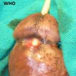
Clinical findings: the patient was alert and hemodynamically stable, without respiratory distress; the recorded World Health Organization performance status was 1. Examination showed marked jaundice, right upper quadrant tenderness without guarding, and moderate ascites. A painful ulceronecrotic umbilical lesion had a surrounding erythematous halo (Figure 1). Several mobile, painless subcutaneous abdominal wall nodules were palpable. No palpable hepatosplenomegaly or peripheral lymphadenopathy was documented.
Timeline: symptoms developed over approximately two months before admission on 3 December 2025. Contrast-enhanced computed tomography and endoscopic ultrasound with pancreatic biopsy were performed on 5 December 2025, two days after admission. The pancreatic biopsy was reported to confirm adenocarcinoma; the date on which the pathology result became available was not recorded. The patient died approximately one month after the biopsy, in early January 2026; the exact date of death was unavailable. These dates and intervals reflect the treating physician's corrected chronology.
Diagnostic assessment
Diagnostic methods: reported admission laboratory values are presented in Table 1. Anemia, lymphopenia, hypoalbuminemia, increased alkaline phosphatase, inflammatory marker elevation, and raised carcinoembryonic antigen and carbohydrate antigen 19-9 (CA 19-9) were reported. The original case-specific laboratory report was not available for independent verification of all units and assay intervals. Total bilirubin was reported as 249 without an established source unit and is retained as such rather than converted. Diagnostic paracentesis yielded clear, straw-colored fluid with a total protein concentration of 30 g/L. Ascitic albumin, a serum-ascites albumin gradient, cell and differential counts, microbiological results, and cytology were unavailable. These data do not establish malignant ascites or exclude infection; the cause of the ascites remained unconfirmed.
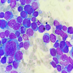
Contrast-enhanced abdominopelvic computed tomography showed a hypoenhancing pancreatic body and tail mass measuring approximately 7.9 x 3.0 cm, with adjacent fat infiltration and splenic artery encasement. Multiple hypodense hepatic lesions were considered suspicious for metastases (Figure 2). Moderate ascites, upper abdominal infiltrative changes, and a hypodense splenic area interpreted as infarction were also described (Figure 3). Endoscopic ultrasound identified a pancreatic body and tail mass and enabled needle biopsy. Histopathological examination of the pancreatic sample was reported to confirm adenocarcinoma. The supplied photomicrograph illustrates the pancreatic sample (Figure 4); the source manuscript describes atypical glandular proliferation in desmoplastic stroma. The treating physician confirmed that the pancreatic biopsy images belonged to this case. No immunohistochemical profile is available for reporting.
Diagnostic challenges: the umbilical lesion was not biopsied, and neither histological nor immunohistochemical confirmation of an umbilical metastasis was available. Original transabdominal ultrasound images were unavailable, so detailed claims attributed to that examination are not used as supporting evidence. Selected computed tomography images and pancreatic biopsy photomicrographs were available, but the complete imaging series and a signed pathology report were not supplied for this revision.
Diagnosis and differential diagnoses: the pancreatic diagnosis was adenocarcinoma according to the treating physician's report of the pancreatic biopsy. Hepatic lesions were radiologically suspicious for metastases. The umbilical lesion was considered clinically suspicious for a Sister Mary Joseph nodule, without tissue confirmation. Alternative explanations for its appearance include an inflammatory or infectious lesion and a primary cutaneous neoplasm; these were not excluded histologically. The other abdominal wall nodules and the ascites also remained unconfirmed as metastatic disease.
Prognostic characteristics: the large pancreatic mass, vascular involvement, suspected hepatic dissemination, ascites, and constitutional symptoms were concerning for advanced disease. The available material does not permit a fully documented pathological stage or establish the mechanism of death.
Therapeutic intervention: diagnostic procedures included paracentesis and endoscopic ultrasound-guided pancreatic biopsy. The treating history indicates that the patient died before tumor-directed treatment. Details of supportive medications, doses, duration, biliary interventions, and changes to treatment were not available and cannot be reconstructed.
Follow-up and outcomes: the treating physician reported death approximately one month after pancreatic biopsy. No serial laboratory or imaging follow-up, formal treatment-response assessment, or documented cause of death was available. Treatment adherence, tolerability, and procedure-related adverse events were not documented; their absence cannot be assumed.
Patient perspective: a patient perspective was not recorded before death and is unavailable.
Informed consent: written informed consent for the publication of this case report and the accompanying images was provided by the patient's brother, as next of kin, because the patient had died. The patient's identity is not disclosed.
Recognition and diagnostic interpretation: the clinical problem in this case was an ulceronecrotic umbilical lesion occurring alongside jaundice, weight loss, ascites, and a pancreatic mass. This combination justified concern for internal malignancy. The nodule remained an important diagnostic clue even though it was not sampled. Published descriptions emphasize examining umbilical lesions carefully and obtaining tissue when appropriate [1,2]. In this patient, pancreatic tissue established the reported primary diagnosis; it did not establish the nature of the separate umbilical lesion.
Comparison with pancreatic cases in the literature: Bai et al. described a 40-year-old man whose erythematous, indurated umbilical lesion was initially treated as eczema. Umbilical biopsy demonstrated adenocarcinoma, while imaging identified a pancreatic neck and body mass with vascular involvement. Dissemination prevented resection, and the patient died four months later after declining chemotherapy [3]. Our patient shared the pancreatic body involvement and vascular encasement, but had a visibly ulceronecrotic lesion, prominent jaundice, and ascites. Crucially, Bai et al. [3] obtained tissue from the umbilicus, whereas the biopsy in our case was pancreatic. Ozaki et al. reported four cases of pancreatic cancer associated with Sister Mary Joseph nodules, all originating in the body and/or tail [4]. The location of our patient's pancreatic primary is consistent with that series. Bolanaki et al. also documented an umbilical nodule as the first sign of pancreatic carcinoma [5]. These reports support considering a pancreatic source when an umbilical lesion accompanies abdominal symptoms, but anatomical similarity does not prove cutaneous metastasis in an individual unbiopsied lesion. Our patient's death approximately one month after pancreatic biopsy documents a short clinical course. It cannot be used to infer that the umbilical lesion caused deterioration or that an alternative treatment would have changed survival. The published cases and this observation differ in diagnostic sampling, extent of documented disease, and management, which limits direct outcome comparison.
Systemic evaluation and interpretation of laboratory findings: when an umbilical lesion raises concern for metastasis, evaluation should connect the skin finding with abdominal examination, imaging, and an appropriate tissue target [1-3]. In this case, computed tomography defined the pancreatic mass and suspicious hepatic lesions, and pancreatic biopsy provided the reported histological diagnosis. A biopsy of the umbilical lesion would have addressed a different unresolved question: whether the skin lesion represented dissemination from that primary. CA 19-9 was elevated, but cholestasis reduces its diagnostic specificity; benign biliary obstruction can also increase its concentration [6]. Neither CA 19-9 nor carcinoembryonic antigen should be used to assign the histology of an unbiopsied skin lesion. The laboratory table describes the reported systemic abnormalities, with explicit uncertainty about source units and intervals, rather than claiming to exclude all differential diagnoses. Similarly, a straw-colored appearance and ascitic total protein of 30 g/L do not supply the missing cytological, microbiological, or cellular findings. The available record leaves both the cause of ascites and its infection status unresolved. The marked jaundice is reported as observed; without the original transabdominal ultrasound or a complete assessment of the biliary tree, the precise mechanism of cholestasis cannot be assigned.
Prognostic implications and limitations: confirmed umbilical metastasis generally accompanies advanced disease and an unfavorable prognosis, although survival varies with the primary cancer and extent of disease [1]. In this case, the pancreatic mass and suspected hepatic spread already indicated a serious prognosis independently of the skin finding. Earlier recognition of a suspicious lesion may facilitate diagnosis and assessment, but this case cannot establish a survival benefit from earlier investigation. The main diagnostic limitation is the lack of umbilical histology. Other limitations include unavailable ascitic-fluid characterization, incomplete case-specific laboratory documentation, selected rather than complete imaging, and absent detailed treatment and follow-up records. The pancreatic diagnosis is based on the treating physician's confirmation and supplied biopsy images; a signed pathology report was not independently available for this revision. These limitations restrict diagnostic certainty at metastatic sites and prevent reconstruction of a complete stage or treatment course.
An ulceronecrotic umbilical lesion accompanying jaundice, abdominal symptoms, or constitutional decline warrants prompt evaluation for internal malignancy. In this patient, pancreatic biopsy was reported to confirm adenocarcinoma, while the unbiopsied umbilical lesion remained clinically suspicious for a Sister Mary Joseph nodule. Imaging, tissue findings, and clinical suspicion should be distinguished explicitly, particularly when documentation of metastatic sites and follow-up is incomplete.
The authors declare no competing interests.
Fatima Zahra Achdami contributed to patient care, provision of the clinical history, and preparation of the case narrative. Individual contributions and final approval by Oussama Nacir and Raouia Daaoufi have not been documented for this revision. Their names and order are retained from the supplied manuscript pending resolution of authorship; supervision or writing contributions are not attributed without supporting information.
Table 1: reported admission laboratory results and comparator intervals
Figure 1: (A,B) the umbilical lesion: an ulceronecrotic center and surrounding erythema are visible; the lesion was clinically suspicious for a Sister Mary Joseph nodule but was not biopsied
Figure 2: selected contrast-enhanced computed tomography images: A) pancreatic body and tail mass, measured at approximately 7.9 x 3.0 cm; B) splenic artery involvement; C) a hypodense hepatic lesion considered suspicious for metastasis; Rred annotations and panel letters are present in the supplied figure
Figure 3: additional contrast-enhanced computed tomography images: A) ascites and upper abdominal infiltrative changes (arrows); B) a hypodense splenic area interpreted as infarction (ellipse). The selected images do not establish the cytological nature of ascites
Figure 4: the pancreatic needle-biopsy material: the source manuscript describes atypical glandular proliferation in desmoplastic stroma, consistent with adenocarcinoma, with hematoxylin and eosin staining; this image relates to the pancreas, not the umbilical lesion; oiginal magnification was not documented
- Gabriele R, Conte M, Egidi F, Borghese M. Umbilical metastases: current viewpoint. World J Surg Oncol. 2005 Feb 21;3(1):13. PubMed | Google Scholar
- Segovis CM, Dyer RB. The "Sister Mary Joseph Nodule". Abdom Radiol (NY). 2017;42:1610-1611. PubMed
- Bai XL, Zhang Q, Masood W, Masood N, Tang Y, Cao CH et al. Sister Mary Joseph's nodule as a first sign of pancreatic cancer. World J Gastroenterol. 2012;18(45):6686-6689. PubMed | Google Scholar
- Ozaki N, Takamori H, Baba H. Sister Mary Joseph's nodule derived from pancreatic cancer. J Hepatobiliary Pancreat Sci. 2011;18(1):119-121. PubMed | Google Scholar
- Bolanaki H, Courcoutsakis N, Kouklakis G, Kakolyris S, Karayiannakis AJ. Sister Mary Joseph's nodule as the first sign of pancreatic carcinoma. J Gastrointest Cancer. 2012;43(Suppl 1):S254-S257. PubMed | Google Scholar
- Ballehaninna UK, Chamberlain RS. The clinical utility of serum CA 19-9 in the diagnosis, prognosis and management of pancreatic adenocarcinoma: an evidence based appraisal. J Gastrointest Oncol. 2012;3(2):105-119. PubMed | Google Scholar
Search

This article authors
On Pubmed
On Google Scholar
Citation [Download]
Navigate this article
Similar articles in
Key words
Tables and figures



Article metrics
Recently from the PAMJ-CM