Pleomorphic adenoma of the buccal mucosa in a 19-year-old woman: an uncommon presentation: a case report
Mainassara Chekaraou Samir, Dandy Mahamadou, Rabe Amani Abdoul Hafizou, Waly Moussa, Habibou Ibrahim Abdoul Majid
Corresponding author: Mainassara Chekaraou Samir, Department of Odontology, Niamey Military Hospital, Niamey, Niger 
Received: 20 Aug 2026 - Accepted: 09 Sep 2026 - Published: 18 Sep 2026
Domain: Oral and Maxillofacial Pathology, Stomatology
Keywords: Pleomorphic adenoma, buccal mucosa, minor salivary gland, young adult, case report
Funding: This work received no specific grant from any funding agency in the public, commercial, or non-profit sectors.
©Mainassara Chekaraou Samir et al. PAMJ Clinical Medicine (ISSN: 2707-2797). This is an Open Access article distributed under the terms of the Creative Commons Attribution International 4.0 License (https://creativecommons.org/licenses/by/4.0/), which permits unrestricted use, distribution, and reproduction in any medium, provided the original work is properly cited.
Cite this article: Mainassara Chekaraou Samir et al. Pleomorphic adenoma of the buccal mucosa in a 19-year-old woman: an uncommon presentation: a case report. PAMJ Clinical Medicine. 2026;22:6. [doi: 10.11604/pamj-cm.2026.22.6.55011]
Available online at: https://www.clinical-medicine.panafrican-med-journal.com//content/article/22/6/full
 PDF in process
Case report 

Pleomorphic adenoma of the buccal mucosa in a 19-year-old woman: an uncommon presentation: a case report
Pleomorphic adenoma of the buccal mucosa in a 19-year-old woman: an uncommon presentation: a case report
 Mainassara Chekaraou Samir1,&, Dandy Mahamadou1, Rabe Amani Abdoul Hafizou1, Waly Moussa2, Habibou Ibrahim Abdoul Majid1
&Corresponding author
Pleomorphic adenoma is the most common benign salivary gland neoplasm, but its occurrence in the minor salivary glands of the buccal mucosa is uncommon. Its nonspecific appearance may mimic other benign or malignant soft-tissue lesions. A 19-year-old woman in good general health presented with a painless right buccal swelling that had progressively enlarged over eight months. Intraoral examination showed a firm, mobile, yellowish-white submucosal nodule measuring approximately 1 × 1.5 cm. The initial differential diagnosis included lipoma. The lesion was excised through an intraoral approach under local anesthesia and submitted for histopathological examination. Microscopy demonstrated benign epithelial and myoepithelial proliferations arranged in lobules and around gland-like spaces within abundant myxoid stroma, without malignant features, establishing the diagnosis of pleomorphic adenoma. Pleomorphic adenoma should be considered in the differential diagnosis of a persistent buccal mass, including in young adults. Histopathological examination is essential, and complete surgical excision followed by long-term surveillance is recommended because recurrence may occur after incomplete removal.

Pleomorphic adenoma is a benign salivary gland tumor characterized by epithelial and myoepithelial differentiation within a morphologically variable stromal background. Although it arises most often in the parotid gland, it can originate from minor salivary glands. In the oral cavity, the palate is the predominant site, whereas buccal mucosal involvement is distinctly uncommon [1-4]. Buccal pleomorphic adenoma usually presents as a painless, slowly enlarging, well-circumscribed submucosal nodule. This nonspecific clinical appearance overlaps with that of lipoma, fibroma, neurogenic tumors, vascular lesions, and other salivary gland neoplasms [1,2]. Histopathological examination is therefore required for definitive diagnosis. We report a pleomorphic adenoma of the right buccal mucosa in a healthy 19-year-old woman and highlight the diagnostic and therapeutic considerations associated with this unusual site and age. This case report was prepared in accordance with the CARE guidelines [5].

Patient information: a 19-year-old woman in good general health was referred for evaluation of a painless swelling of the right buccal region. She had first noticed the lesion approximately eight months before consultation and reported progressive enlargement. Her medical history was unremarkable, and no previous intervention for the lesion was reported.
Clinical findings: extraoral examination was unremarkable, with no clinically palpable regional lymphadenopathy. Intraoral examination revealed a firm, mobile, yellowish-white submucosal lesion of the right buccal mucosa measuring approximately 1 × 1.5 cm. The overlying mucosa was intact ( Figure 1).
Timeline: eight months before consultation, the patient first noticed a painless swelling of the right buccal mucosa, which gradually increased in size. At presentation, examination identified a firm, mobile submucosal nodule measuring approximately 1 × 1.5 cm.
Diagnostic assessment: the clinical appearance suggested a benign soft-tissue lesion, and the initial differential diagnosis included lipoma. Other diagnostic considerations for a buccal submucosal mass include fibroma, neurogenic tumor, vascular lesion, and benign or low-grade malignant minor salivary gland neoplasm.
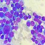
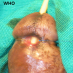
Therapeutic intervention: the lesion was approached intraorally and completely excised under local anesthesia. Careful dissection was performed while preserving Stensen's duct; the exposed lesion before removal is shown in Figure 2. Gross examination of the excised specimen showed a firm, homogeneous, whitish fragment with a fibrous appearance and a greatest dimension of 1.5 cm The specimen was submitted for histopathological examination. Histologically, the specimen contained a benign biphasic salivary gland tumor composed of epithelial and myoepithelial proliferations. Epithelial cells formed solid lobules and groups around gland-like spaces, while the myoepithelial component comprised regular spindle-shaped cells. The stroma was abundant and myxoid, without cartilaginous or osseous differentiation. No malignant features were identified. These findings established the diagnosis of pleomorphic adenoma ( Figure 3).
Follow-up and outcomes: the postoperative course was straightforward. One-year follow-up revealed no recurrence.
Patient perspective: the patient stated: "I am satisfied with the treatment and no longer feel any discomfort in my cheek."
Informed consent: verbal informed consent was obtained from the patient for publication of this case report and the accompanying clinical, operative, and histopathological images.
Pleomorphic adenoma is the most frequent benign salivary gland neoplasm, but minor salivary gland involvement accounts for only a minority of cases. Among intraoral sites, the buccal mucosa is affected much less often than the palate [1-4]. The present case is additionally notable because the patient was 19 years old; pleomorphic adenoma is encountered more often in middle adulthood, although cases in adolescents and young adults have been reported [3,6,7]. Dalati and Hussein described a cheek pleomorphic adenoma in a 17-year-old girl, confirming that this tumor can occur during adolescence [6]. The usual clinical presentation is an indolent, painless, firm, and mobile submucosal mass covered by intact mucosa. Our patient’s right buccal lesion measured 1 × 1.5 cm after an eight-month course. Clinical findings alone, however, are not specific. The differential diagnosis includes fibroma, lipoma, neurogenic tumors, vascular lesions, nodular fasciitis, and benign or malignant minor salivary gland tumors. Rapid growth, pain, ulceration, fixation, sensory disturbance, or regional lymphadenopathy should raise suspicion of malignancy. Histologically, pleomorphic adenoma displays epithelial and myoepithelial cells arranged in ducts, nests, cords, or sheets within variably myxoid, chondromyxoid, or hyalinized stroma [8]. In the present case, epithelial cells formed solid lobules and groups around gland-like spaces, while regular spindle-shaped myoepithelial cells were embedded in abundant myxoid stroma. The absence of cartilaginous or osseous differentiation does not exclude the diagnosis because the relative proportions and character of the stromal elements vary. No malignant feature was identified.
The recommended treatment of a pleomorphic adenoma arising from a minor salivary gland is surgical excision without capsular rupture, with an adequate margin when anatomically feasible [1-3]. Simple enucleation should be avoided because microscopic pseudopodia, satellite nodules, and tumor spillage may predispose to recurrence. In the buccal region, the surgeon must consider the parotid duct, buccal branches of the facial nerve, and adjacent vascular structures. In this patient, an intraoral approach under local anesthesia was feasible because the lesion was small and accessible. Although no recurrence was observed during one year of follow-up, longer surveillance remains warranted because recurrence may be delayed, especially after incomplete removal. Malignant transformation into carcinoma ex pleomorphic adenoma is uncommon and is associated mainly with longstanding or recurrent tumors.
Pleomorphic adenoma should be included in the differential diagnosis of a persistent buccal submucosal mass, even in a young adult. Because clinical findings are nonspecific, histopathological examination is indispensable. Careful excision without capsular rupture and long-term follow-up are central to limiting recurrence. The key clinical lesson is that an apparently innocuous cheek nodule may represent a minor salivary gland neoplasm.
The authors declare no competing interests.
Patient management: Mainassara Chekaraou Samir, Rabe Amani Abdoul Hafizou, Habibou Ibrahim Abdoul Majid, and Dandy Mahamadou; data collection: Waly Moussa; manuscript drafting: Mainassara Chekaraou Samir, Rabe Amani Abdoul Hafizou, Habibou Ibrahim Abdoul Majid, and Dandy Mahamadou; manuscript revision: Mainassara Chekaraou Samir. All authors read and approved the final version of the manuscript.
Figure 1: preoperative intraoral appearance of the right buccal submucosal swelling, measuring approximately 1 × 1.5 cm
Figure 2: intraoperative view after dissection, showing the exposed, well-circumscribed right buccal lesion before removal
Figure 3: showing epithelial and myoepithelial tumor components within abundant myxoid stroma, consistent with pleomorphic adenoma
- Júnior EC, Dias IJ, Alves PM, Nonaka CF, Gomes DQ, Pereira JV. Pleomorphic adenoma of buccal mucosa: a case report and review of the literature. Stomatologija. 2020;22(3):92-96. Google Scholar
- Periasamy S, Manoharan A, Garg H, Kumar SP. Pleomorphic adenoma of the cheek: a case report. Cureus. 2019 Aug 3;11(8):e5312. PubMed | Google Scholar
- AlWatban Z, AlAnsari H, AlAseeri D, AlJawder A, Janahi W. Pleomorphic adenoma of the oral cavity: systematic review. Indian J Otolaryngol Head Neck Surg. 2023 Dec;75(4):3288-3304. PubMed | Google Scholar
- Lianou AD, Zarachi A, Derka S, Sioka C, Ragkos NG, Liontos A et al. Pleomorphic adenoma of minor salivary glands: a rare case report. Maedica (Bucur). 2024;19(2):434-438. PubMed | Google Scholar
- Gagnier JJ, Kienle G, Altman DG, Moher D, Sox H, Riley D et al. The CARE guidelines: consensus-based clinical case reporting guideline development. J Clin Epidemiol. 2014;67(1):46-51. PubMed | Google Scholar
- Dalati T, Hussein MR. Juvenile pleomorphic adenoma of the cheek: a case report and review of literature. Diagn Pathol. 2009 Sep 22;4:32. PubMed | Google Scholar
- Jagadishkumar K, Anilkumar MG, Krishna Murthy DS, Puneeth HK. Pleomorphic adenoma of the cheek in a child: a case report. Dent Res J (Isfahan). 2014;11(4):522-524. PubMed | Google Scholar
- Triantafyllou A, Thompson LD, Devaney KO, Bell D, Hunt JL, Rinaldo A et al. Functional histology of salivary gland pleomorphic adenoma: an appraisal. Head Neck Pathol. 2015;9(3):387-404. PubMed | Google Scholar
Search

This article authors
On Pubmed
On Google Scholar
Citation [Download]
Navigate this article
Similar articles in
Key words
Tables and figures


Article metrics
Recently from the PAMJ-CM