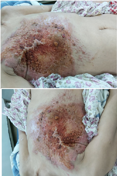
Radiation therapy has been one of the pillars of cancer therapy for many years. Many technological advances have been made to improve radiation delivery to the targeted area while reducing damage to the surrounding normal tissue. The spectrum of late radiation-induced injuries is quite heterogeneous but the injuries are usually benign. They may manifest themselves as breast edema, fibrosis, telangiectasia, pulmonary fibrosis, chronic ulceration, rib fractures, fat necrosis or osteonecrosis. We report the case of a 55-year-old woman who presented to the Neurosurgery Department for management of hydrocephalus with a posterior fossa tumor. The patient had a past history of a multifocal adenocarcinoma of the both breast, which were resected 13 years earlier. Further treatment included chemotherapy with six cycles of doxorubicin and cyclophosphamide in combination with cobalt radiation with a total dose of 60 Gy, followed by tamoxifen for a total of 4 years. The first local signs were observed two years after the radiation treatment with the loss of irradiated skin elasticity followed by increasing induration and telangiectasia. Over the one year before presentation, the mass had significantly increased in size and become increasingly painful, restricting the patient psychologically and functionally in her daily activities. Local examination revealed a subcutaneous fat necrosis of the right breast, which had been progressively growing over a period of 02 years (A).
Corresponding Author
Hassan Baallal, Department of Neurosurgery, Avicenne Military Teaching Hospital, University Caddi Ayyad, Marrakech, Morocco (baallalnch@gmail.com)