Intact amniotic sac prolapsing through a lower uterine segment rupture: a striking intraoperative image
Farha Fareed Khan, Mohammed Faizan Ulla Khan
Corresponding author: Farha Fareed Khan, Obstetrics and Gynaecology, Datta Meghe Institute of Medical Sciences, Sawangi (Meghe), Wardha, Maharashtra, India 
Received: 02 Jul 2026 - Accepted: 07 Jul 2026 - Published: 12 Aug 2026
Domain: Gynecology, Obstetrics and gynecology
Keywords: Abruptio placentae, uterine scar dehiscence, lower-segment caesarean sections, exploratory laparotomy
Funding: This work received no specific grant from any funding agency in the public, commercial, or non-profit sectors.
©Farha Fareed Khan et al. PAMJ Clinical Medicine (ISSN: 2707-2797). This is an Open Access article distributed under the terms of the Creative Commons Attribution International 4.0 License (https://creativecommons.org/licenses/by/4.0/), which permits unrestricted use, distribution, and reproduction in any medium, provided the original work is properly cited.
Cite this article: Farha Fareed Khan et al. Intact amniotic sac prolapsing through a lower uterine segment rupture: a striking intraoperative image. PAMJ Clinical Medicine. 2026;21:23. [doi: 10.11604/pamj-cm.2026.21.23.54303]
Available online at: https://www.clinical-medicine.panafrican-med-journal.com//content/article/21/23/full
Images in clinical medicine 

Intact amniotic sac prolapsing through a lower uterine segment rupture: a striking intraoperative image
Intact amniotic sac prolapsing through a lower uterine segment rupture: a striking intraoperative image
Farha Fareed Khan1,&,  Mohammed Faizan Ulla Khan2
&Corresponding author

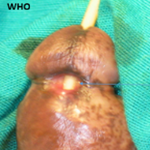
Uterine rupture, though a well-recognized obstetric catastrophe, rarely presents itself with the intraoperative finding of an intact, prolapsed amniotic sac through the uterine defect, an exceptionally uncommon finding reported only in isolated case reports. A 28-year-old woman (G3P2) with two prior lower-segment caesarean sections (LSCS) presented at 34 weeks of gestation with sudden-onset severe abdominal pain and absent fetal movements since eight hours. On examination, patient was pale, in haemorrhagic shock (pulse 112 beats per minute; blood pressure 88/60 mmHg), with a tender abdomen and absent fetal heart sounds. Ultrasonography revealed an empty uterine cavity, intrauterine fetal demise, a defect in the anterior lower uterine wall, and free intraperitoneal fluid. Following resuscitation, emergency laparotomy was performed under general anaesthesia. Intra-operatively, a 6-cm longitudinal rupture was identified along the prior caesarean scar of the lower uterine segment. The striking intraoperative finding-depicted in the figure-was the complete prolapse of an intact, bulging amniotic sac through this uterine defect into the peritoneal cavity. Approximately 800 mL of haemoperitoneum was evacuated. The membranes were incised, and a stillborn male fetus (weight 2.0 kg) was delivered. The uterine defect was repaired in two interrupted layers, and bilateral tubal ligation was performed simultaneously. The patient had an uneventful postoperative recovery and was discharged on postoperative day four.
Figure 1: intraoperative view showing intact amniotic sac prolapsing through a lower uterine segment rupture along the prior caesarean scar
Search

This article authors
On Pubmed
On Google Scholar
Citation [Download]
Navigate this article
Similar articles in
Key words
Tables and figures

Article metrics
Recently from the PAMJ-CM