Diagnostic and therapeutic considerations in apical and mid-cavity hypertrophic cardiomyopathy: a case series on three Ghanaian patients
Aba Folson, Harold Ayetey, Francis Agyekum, Kwabena Oteng Agyapong
Corresponding author: Aba Folson, Department of Internal Medicine, School of Medicine, University of Health and Allied Sciences, Ho, Ghana 
Received: 09 Mar 2026 - Accepted: 13 May 2026 - Published: 14 Jul 2026
Domain: Cardiology
Keywords: Hypertrophic, cardiomyopathy, Ghanaian
Funding: This work received no specific grant from any funding agency in the public, commercial, or not-for-profit sectors.
©Aba Folson et al. PAMJ Clinical Medicine (ISSN: 2707-2797). This is an Open Access article distributed under the terms of the Creative Commons Attribution International 4.0 License (https://creativecommons.org/licenses/by/4.0/), which permits unrestricted use, distribution, and reproduction in any medium, provided the original work is properly cited.
Cite this article: Aba Folson et al. Diagnostic and therapeutic considerations in apical and mid-cavity hypertrophic cardiomyopathy: a case series on three Ghanaian patients. PAMJ Clinical Medicine. 2026;21:19. [doi: 10.11604/pamj-cm.2026.21.19.52083]
Available online at: https://www.clinical-medicine.panafrican-med-journal.com//content/article/21/19/full
 PDF in process
Case series 

Diagnostic and therapeutic considerations in apical and mid-cavity hypertrophic cardiomyopathy: a case series on three Ghanaian patients
Diagnostic and therapeutic considerations in apical and mid-cavity hypertrophic cardiomyopathy: a case series on three Ghanaian patients
Aba Folson1,2,&, Harold Ayetey3,  Francis Agyekum4, Kwabena Oteng Agyapong2
&Corresponding author
Hypertrophic cardiomyopathy (HCM) is a genetic cardiac muscle disorder that typically results in asymmetric hypertrophy of ventricular muscle and may cause sudden cardiac death. Apical and mid-cavity hypertrophic cardiomyopathy are less common phenotypes of this condition inherited in an autosomal dominant manner. A low index of suspicion may influence the perceived rarity and varied outcomes among African populations, with associated confounders contributing to misdiagnosis. Inaccessibility and unaffordability of appropriate diagnostic and therapeutic tools remain relevant in assessing such patients. We explore these factors in this case series on three Ghanaian patients with varying phenotypes of HCM. Two middle-aged male patients both with apical HCM are described. One patient, found to be hypertensive as well as engaging in isometric exercises, was asymptomatic for heart failure or ischaemia but complained of intermittent palpitations. The third patient presented with recurrent palpitations with no syncope and was found to have frequent episodes of non-sustained ventricular tachycardia. The third patient was a middle-aged female with left ventricular mid-cavity obstruction and apical aneurysm. All three patients had diagnostic and/or therapeutic challenges.

Hypertrophic cardiomyopathy is a genetic disorder with several morphologic variants resulting from myocyte disarray and fibrosis with segmental hypertrophy [1]. The commonest form is asymmetrical septal hypertrophy; less common forms are apical hypertrophic cardiomyopathy and mid-cavity obstructive cardiomyopathy (MVO) [2]. Generally, HCM is defined as a left ventricular wall thickness of 15 mm or more in any segment with no loading condition. Alternatively, the diagnosis can be made with dimensions less than 15 mm but with other consistent clinical and imaging/pathological features such as a family history of HCM, asymmetric distribution of the thickening, myocardial fibrosis on CMR or distinctive ECG changes [3]. Apical hypertrophic cardiomyopathy (ApHCM), an atypical and rare phenotype of hypertrophic cardiomyopathy, predominantly affects the apex of the left ventricle. It is more common in the Asian population (affecting about 25% of persons with hypertrophic cardiomyopathy) than in other parts of the world (incidence reported at 3- 11% of all hypertrophic cardiomyopathy cases). The peak incidence occurs in mid-life (especially in the 4th decade) with about 6% of patients reporting a positive family history of ApHCM [4]. Owing to its frequently non-specific presentation both clinically and in imaging, diagnosis of HCM is often delayed and further confounded by the presence of other causes of left ventricular hypertrophy such as hypertension, diabetes mellitus [5] and long-standing exercise [6]. Mid-cavity obstructive cardiomyopathy (MVO) is the least common form of HCM with very few studies conducted on it and none documented among Africans to date. In one study on athletes, 43 out of 423 patients with HCM had MVO and left ventricular outflow tract obstruction [7]. Of these, 62.5% were females and 26.5% had apical aneurysms, confirmed after coronary artery disease was actively screened for and excluded. We present three cases of HCM two of whom have ApHCM and a third with features in keeping with severe MVO. This report seeks to explore the diagnostic challenges for such patients in Ghana.

Case 1
A 57-year-old male patient presented for an echocardiogram after experiencing intermittent palpitations for a month. Despite being generally well (with no dyspnoea, chest pain, or syncope), he was experiencing worsening palpitations during the day and becoming more fatigued with activities he was previously able to undertake without symptoms. The patient developed hypertension four years prior on candesartan 16 mg nocte and type two diabetes of two years duration on metformin 500 mg twice a day. He was on 40 mg of atorvastatin at night for dyslipidemia. The patient has been involved in weightlifting and bariatric exercises for the past decade, attending the gym twice weekly and participating in bodybuilding activities like dumbbell shoulder press and bench press exercises. He has no history of using anabolic steroids, alcohol, nicotine, or illicit drugs. Physical examination showed a tall, muscular male with a body mass index (BMI) of 38kg/m2 with no anemia or peripheral edema. His pulse rate was 78 beats per minute and was regular at the time and the blood pressure was 156/98 mmHg. His jugular venous pulse was not elevated and the apex beat was heaving but undisplaced. Heart sounds were normal and there was no murmur heard. No fine bi-basal crepitations were auscultated and all other systems were unremarkable.
Case 2
A 56-year-old man presented with occasional chest discomfort and recurrent episodes of palpitations but no syncope or presyncope. He had a history of hypertension, dyslipidaemia and cervical spondylosis. He had no family history of sudden cardiac death. He was a non-smoker and drank less than 14 units of alcohol a week. He was on amlodipine 5mg and carvedilol 12.5mg od for his blood pressure and palpitations which were thought to be due to occasional premature ventricular ectopic beats by his primary physician though the ECG did not indicate antiarrhythmia. At the cardiology clinic, he was apyrexial with a blood pressure of 129/78mmHg, a pulse of 61bpm and oxygen saturation of 98% on room air. Apart from appearing obese (BMI 30kg/m2), his clinical examination was essentially normal. His routine labs, full blood count (FBC), blood urea and electrolytes with creatinine (BUE/Cr), liver function test (LFT) and glycated hemoglobin (HbA1c) were all unremarkable. His low-density lipoprotein cholesterol was however elevated at 4.2mmol/L with a total cholesterol of 5.78mmol/L.
Case 3
A 61-year-old female, hypertensive for the past 6 years presented to the cardiac clinic with non-exertional central chest pain for the past two weeks. She also had intermittent palpitations with occasional associated dizziness. She had no history of syncope or difficulty breathing. Current medications included Amlodipine 5 mg and Bisoprolol 5 mg daily since being diagnosed hypertensive. She worked as a trader and only occasionally drank alcohol. There was no family history of sickle cell disease (SCD) or HCM. Physical examination showed an overweight female with, a BMI of 27.5kg/m2 in no respiratory distress. She had no pallor or pedal oedema. Her pulse rate was 88/min and was regular with normal heart sounds. There was a grade 3 diastolic murmur over an undisplaced apex with a blood pressure of 138/78 mmHg. There was no evidence of fluid overload and the gastrointestinal and central nervous systems were normal.
Case 1
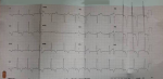
The electrocardiogram (ECG) done, (Figure 1), indicated voltage criteria for left ventricular hypertrophy (LVH) and widespread, asymmetrical T wave inversions that were very deep and prominent in the anterolateral leads. At the time, he had no arrhythmia noted on the ECG and a subsequent 24-hour Holter recording showed intermittent premature ventricular complexes (3.4% of the total beat count) with no bigeminy, trigeminy, episodes of ventricular tachycardia or pauses greater than 2000ms. There were no premature atrial beats, supraventricular tachycardia or periods of atrial fibrillation noted. His haemoglobin was 14.6g/dl and he had an eGFR of 82, a low-density lipoprotein (LDL) cholesterol level of 3.0mmol/l and a chest X-ray with no evidence of pulmonary oedema. His cardiothoracic ratio (CTR) was 0.5 and glycated haemoglobin was 6.2%. The echocardiography revealed mild left atrial dilatation. There was LVH limited to the mid cavity and more severe in the apical region (2.1cm). The myocardium had a speckled appearance. The estimated left ventricular mass indexed to the body surface area was 132g/m2 with grade one diastolic dysfunction and normal left atrial filling pressures. The LV ejection fraction was 75% and the stroke volume was 86ml. There was no evidence of pulmonary hypertension. A diagnosis of apical hypertrophic cardiomyopathy was considered alongside hypertensive heart disease with the possible contribution of the athlete´s heart to the observed hypertrophy.
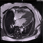
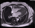
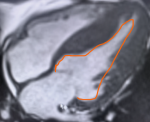
A cardiac magnetic resonance imaging (MRI) was done which showed normal LV volumes and ejection fraction (76%), hyperdynamic contractility with apical occlusion, increased LV apical myocardial thickness (max 2.3 cm apical anteroseptum vs 1.8cm apical lateral wall) with apical insertion of hypertrophied posterior papillary muscle. There was no left ventricular outflow tract obstruction (LVOT) obstruction, systolic anterior motion of the septal wall (SAM) or regional wall motion abnormality seen (RWMA). No systolic cavity occlusion or aneurysm formation was observed. In the gadolinium study, the early phase demonstrated no LV/RV thrombus whereas the late phase showed patchy enhancement of hypertrophied apical LV segments. The pattern of hypertrophy and patchy fibrosis of the asymmetrically thickened LV wall was consistent with ApHCM (Figure 2). The patient's first-degree relatives, including his brother and sister, underwent screening ECG and echocardiography, but no findings indicated HCM. He is currently being treated with candesartan 16 mg daily, bisoprolol 5 mg daily, metformin 500mg twice a day and 40 mg of atorvastatin at night. A repeat 24-hour Holter recording done 2 months into therapy showed the proportion of PVCs to be less than 1%. He had no chest pain or palpitations and easy fatigue has improved. His last measured blood pressure was 118/78 mmHg. The patient has been advised to gradually reduce the intensity of bariatric exercises with the view to stopping themcompletely. He will be followed up at the cardiology outpatient clinic with periodic ECG and echocardiography.
Case 2
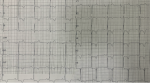
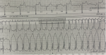
His ECG showed LVH with widespread T wave inversion (Figure 3), while a 24-hour Holter showed several multifocal ventricular ectopic beats with two runs of non-sustained VT, the longest lasting 7.5 seconds (Figure 4). An echocardiogram showed severe asymmetric LVH with mid and apical obstruction, a dilated LA (40mm) and grade 3 diastolic dysfunction with a normal ejection fraction (65%) and no regional wall motion abnormality. A cardiac MRI revealed a spade-shaped LV cavity with normal systolic function, complete systolic apical cavity obliteration and severe asymmetrical LVH with patchy late gadolinium enhancement in the hypertrophied mid-cavity to apical segments. No LVOT obstruction or SAM was observed, all consistent with apical HCM (Figure 5). First-degree relatives were screened by echocardiography with no findings to indicate HCM. His 5-year risk of sudden cardiac death was assessed with the HCM Risk SCD calculator and deemed to be intermediate to high at approximately 5% based on his age of 57years, maximal wall thickness of 28mm, left atrial size of 40mm, LVOT gradient of 4 mmHg, presence of non-sustained VT, and absence of family history of SCD or unexplained syncope. The patient was referred outside of Ghana to a cardiac electrophysiologist and an ICD was implanted based on an HCM sudden cardiac risk score (HCM risk-SCD) of between 4 and 5.13% (intermediate risk group) for which ICDs can be considered. He remains on amlodipine 5mg, carvedilol 12.5mg, and rosuvastatin 10mg daily and is doing well.
Case 3
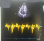
The patient had an ECG done which indicated no left ventricular hypertrophy but widespread inverted T waves. On the echocardiogram, there was a thinned-out LV apex with hypertrophied mid-segment septal and lateral walls that apposed during systole depicting a somewhat hourglass outline. There was a high flow velocity of 4.1m/s with a pressure gradient of 68 mmHg (Figure 6) in the mid cavity of the left ventricle. Left ventricle ventricular ejection fraction of 78% with a reduced stroke volume of 62ml and grade 1 diastolic dysfunction was present. There was no evidence of pulmonary hypertension noted and no thrombus was seen in the LV apex. The 24-hour Holter showed occasional premature ventricular complexes (3.2% of the total beat count) with a run of non-sustained ventricular tachycardia noted. The treadmill exercise test was negative for inducible ischaemia and there was no worsening of arrhythmia during or immediately after the exercise. A diagnostic coronary angiogram did not show significant epicardial vessel disease.
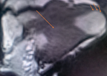
The cardiac MRI showed normal LV volumes and ejection fraction (66%). Hyperdynamic contraction and increased LV myocardial thickness (max 2.5cm mid-inferoseptal vs 1.8cm mid-lateral wall) with no inflammation or edema noted. Apically inserted papillary muscles were noted alongside mid-cavity occlusion in systole with an apical aneurysm measuring 3x3.6cm. No LVOT obstruction was present, but systolic anterior motion of the anterior mitral valve leaflet (SAM) was noted. There was no regional wall motion abnormality seen. The patchy fibrosis of the asymmetrically thickened LV wall was most consistent with MVO (Figure 7). Following the diagnosis, the patient's three sons were all screened with ECG and echocardiography. One of them had mild concentric left ventricular hypertrophy due to hypertensive heart disease whilst the others had no muscle mass abnormalities. The patient and son will be followed up with yearly echocardiography. Counselling on the risk of SCD from life-threatening arrhythmias, the possibility of forming intracardiac thrombi and the development of heart failure as well as the need for long-term remote monitoring and possibly the insertion of an ICD has been done. Unfortunately, further electrophysiologic assessment and intervention are neither available nor financially accessible to this patient. She is currently on Telmisartan 80 mg daily, carvedilol 6.25mg daily and Rivaroxaban 20 mg daily.
Hypertrophic cardiomyopathy (HCM) presents with segmental hypertrophy, with the pattern of this distribution resulting in several phenotypes. Less common forms such as ApHCM and MVO, which have fairly distinctive patterns of distribution, may still present a diagnostic challenge. In Africa, where the documentation and management of these subtypes is uncommon, factors that may contribute to this include the lack of appropriate and almost basic diagnostic tools such as echocardiography, the inadequacy of trained personnel, the impact of confounding factors and less than optimal care due to economic and infrastructural constraints. The ECG may present with ST and T wave abnormalities (>10mm deep T wave inversions in inferolateral leads) as the first indication of HCM and ApHCM [8]. Commonly, this may be misdiagnosed as coronary artery disease or LVH unless followed up with other imaging modalities and biochemical tests to exclude these differential diagnoses. The lack of widespread availability of echocardiography, a basic cardiovascular imaging tool in LMICs limits its applicability in the screening and detection of all forms of HCM. Additionally, several confounding causes of LVH including hypertension and intense exercise, contributing to misdiagnosis may be excluded with some level of certainty if imaging by echocardiography is accessible. Adebayo et al. describe the presence of 4 geometric phenotypes of LVH among Nigerian patients with hypertension all of which could potentially camouflage HCM [9]. Speckle-tracking echocardiography may help distinguish HCM from LVH in hypertension as the former is associated with a greater increase in global longitudinal strain and generally, the concomitant presence of cardiovascular risk factors is associated with more severe HCM phenotypes [10]. It is important to recognise that increased wall thickness in the setting of a reduced LV chamber dimension is highly unlikely to be explained solely by exercise-induced physiological adaptation and should always initiate a search for other causes, especially among athletes [11]. The left ventricular internal diameter in diastole has been cited as a discriminator of HCM and the athlete's heart and septal wall thickness of 14mm in males and 11 mm in female athletes of African descent may be considered physiological [12].
Diagnostic ambiguity associated with the use of 2D echocardiography may be overcome with the use of cardiac magnetic resonance imaging (CMR) where less typical segmental hypertrophy such as is seen in ApHCM and MVO may be detected by the presence of segmental fibrosis and late gadolinium enhancement [13]. Mid-cavity obstructive cardiomyopathy (MVO) variants tend to be more symptomatic at presentation with the occurrence of arrhythmias, in particular, being irrespective of the presence or absence of aneurysms [14]. It is thought that arrhythmogenesis is initiated and sustained by the fibrotic segments acting as substrates which function independently of severe remodelled states such as aneurysms and the consideration of primary prevention of arrhythmias and their sequelae is paramount in the management of such patients. Atrial fibrillation (AF) remains the most prevalent arrhythmia among patients with HCM and was recorded to be as high as 22.45% in one systematic review [15]. Major guideline recommendations encourage the implantation of an ICD in patients above the age of 16 years with at least an intermediate risk of SCD and any of the following: significant late gadolinium enhancement on CMR, LV ejection fraction <50%, abnormal BP response during exercise testing, the presence of an apical aneurysm or the detection of a genetic mutation of the sarcomere [16]. Pharmacologic treatment of VT in HCM includes the use of B-blockers or amiodarone in primary and secondary prevention of lethal ventricular arrhythmias [17]. These pharmacological options, though less effective than implantation of ICD, may be a viable option in LMICs, where device therapy is largely unattainable to those who need it.
Aneurysms in HCM have been theorised to be a result of ischaemia from extravascular compression of coronary arteries leading to chronic hypoxia and ischaemia. Some authors have proposed that MVO with apical aneurysm be recognised as a form of arrhythmogenic cardiomyopathy on account of the significantly increased risk of fatal and nonfatal arrhythmias following the recognition of the aneurysmal rim as the primary substrate for monomorphic VT [17]. Left ventricular apical aneurysms occur in 2 to 5% of HCM and present as a region of akinesia or dyskinesia following scarring. Early surgical interventions to aid in the relief of the pressure gradient within the ventricle such as transapical myectomy and surgical cryoablation are known to help reduce the risk of developing aneurysms [18]. There is an established association between the presence of apical aneurysms in HCM and thromboembolic events and the risk is higher in moderate to large aneurysms though they may occur in smaller ones [19]. Full anticoagulation with re-evaluation after three months following the diagnosis of an LV aneurysm with a thrombus with either a direct oral anticoagulant (DOACs) or warfarin is however recommended [20].
Limitations: this study is a case series and may not be generalizable.
Hypertrophic cardiomyopathy (HCM) with its varying phenotypes is very easily misdiagnosed and mismanaged. Less common variants such as ApHCM and MVO have been under-reported in general and the factors for their under detection, especially in Africa have been explored. Echocardiography remains an invaluable means of early detection of index cases and screening of relatives. The impact of physiological conditions such as exercise and pathologic cardiovascular risk factors such as hypertension however often necessitate advanced imaging techniques to confirm the diagnosis. The detection and optimal management of complications such as aneurysms, thromboembolic phenomena, and particularly fatal and non-fatal arrhythmias, impact survival greatly. Unfortunately accessing these services remains a distant illusion for most patients in Ghana.
What is known about this topic
- Apical and mid-cavity variants if hypertrophic cardiomyopathy is uncommon among African populations;
- Diagnostic delays are common in Africa due to challenges in advanced diagnostic tools.
What this study adds
- This series reminds practitioners in our part of the world that atypical forms of HCM may occur among our population, though they are largely considered rare;
- This study highlights the challenges in the diagnosis of HCM in the setting of confounders such as hypertension and engagement in bariatric exercise.
The authors declare no competing interests.
Aba Folson: conceptualization of the series; writing of original draft, review and editing; case management. Harold Ayetey: conceptualization of the series, review and editing of the write-up; imaging; case management. Francis Agyekum: conceptualization review and editing of the write-up. Kwabena Oteng Agyapong: Conceptualization; review and editing of the write-up. All the authors have read and approved the final version of this manuscript.
The authors would like to thank the patients for their cooperation during the assessment and writing of this report.
Figure 1: electrocardiogram showing voltage criterion left ventricular hypertrophy and widespread deep inverted T waves
Figure 2: severe apical hypertrophy with late-phase patchy fibrosis during the cardiac MRI gadolinium study
Figure 3: ECG showing widespread T wave inversions and left ventricular hypertrophy
Figure 4: 24-hour Holter showing a run of non-sustained ventricular tachycardia
Figure 5: cardiac MRI depicting a spade-shaped ventricular cavity with mid and apical hypertrophy
Figure 6: echocardiographic continuous wave spectrum image showing high LV mid-cavity velocity
Figure 7: cardiac MRI showing severe mid-cavity obstruction and apical aneurysm with thinned-out left ventricular apical wall resulting in an hour-glass cavity
- Kim EK, Lee SC, Hwang JW, Chang SA, Park SJ, On YK et al. Differences in apical and non-apical types of hypertrophic cardiomyopathy: a prospective analysis of clinical, echocardiographic, and cardiac magnetic resonance findings and outcome from 350 patients. Eur Heart J Cardiovasc Imaging. 2016 Jun;17(6):678-86. PubMed | Google Scholar
- Gersh BJ, Maron BJ, Bonow RO, Dearani JA, Fifer MA, Link MS et al. ACCF/AHA guideline for the diagnosis and treatment of hypertrophic cardiomyopathy: a report of the American College of Cardiology Foundation/American Heart Association Task Force on Practice Guidelines. Circulation. 2011 Dec 13;124(24):e783-831. PubMed | Google Scholar
- Parato VM, Olivotto I, Maron MS, Nanda NC, Pandian NG. Left ventricular apex involvement in hypertrophic cardiomyopathy. Echocardiography. 2015 Oct;32(10):1575-80. PubMed | Google Scholar
- Eriksson MJ, Sonnenberg B, Woo A, Rakowski P, Parker TG, Wigle ED et al. Long-term outcome in patients with apical hypertrophic cardiomyopathy. J Am Coll Cardiol. 2002 Feb 20;39(4):638-45. PubMed | Google Scholar
- Kubo T, Kitaoka H, Okawa M, Hirota T, Hoshikawa E, Hayato K et al. Clinical profiles of hypertrophic cardiomyopathy with apical phenotype: comparison of pure-apical form and distal-dominant form. Circ J. 2009 Dec;73(12):2330-6. PubMed | Google Scholar
- Li J, Owusu IK, Geng Q, Folson AA, Zheng Z, Adu-Boakye Y et al. Cardiometabolic risk factors and preclinical target organ damage among adults in Ghana: findings from a national study. J Am Heart Assoc. 2020 Dec 15;9(24):e017492. PubMed | Google Scholar
- SSheikh N, Papadakis M, Schnell F, Panoulas V, Malhotra A, Wilson M et al. Clinical profile of athletes with hypertrophic cardiomyopathy. Circ Cardiovasc Imaging. 2015 Jul;8(7):e003454. PubMed | Google Scholar
- Derosa Giuseppe, Maffioli Pamela. Assessment and management of left ventricular hypertrophy in type 2 diabetes patients with high blood pressure. Expert Rev Cardiovasc Ther. 2013 Jun;11(6):719-28. PubMed | Google Scholar
- Adebayo RA, Bamikole OJ, Balogun MO, Akintomide AO, Adeyeye VO, Bisiriyu LA et al. Echocardiographic assessment of left ventricular geometric patterns in hypertensive patients in Nigeria. Clin Med Insights Cardiol. 2013 Oct 23:7:161-7. PubMed | Google Scholar
- de la Rosa A, Shah M, Shiota T, Siegel R, Rader F. Comparing echocardiographic characteristics in genotype positive-phenotype positive hypertrophic cardiomyopathy and hypertensive left ventricular hypertrophy. Eur Heart J Cardiovasc Imaging. 2022 Feb 22;23(3):340-348. PubMed | Google Scholar
- Brosnan Mark J, Rakhit Dinesh. Differentiating athlete's heart from cardiomyopathies-the left side. Heart Lung Circ. 2018 Sep 1;27(9):1052-62. Google Scholar
- Basavarajaiah S, Boraita A, Whyte G, Wilson M, Carby L, Shah A et al. Ethnic differences in left ventricular remodeling in highly-trained athletes: relevance to differentiating physiologic left ventricular hypertrophy from hypertrophic cardiomyopathy. J Am Coll Cardiol. 2008 Jun 10;51(23):2256-62. PubMed | Google Scholar
- Quarta G, Aquaro GD, Pedrotti P, Pontone G, Dellegrottaglie S, Iacovoni A et al. Cardiovascular magnetic resonance imaging in hypertrophic cardiomyopathy: the importance of clinical context. Eur Heart J Cardiovasc Imaging. 2018 Jun 1;19(6):601-610. PubMed | Google Scholar
- Minami Y, Kajimoto K, Terajima Y, Yashiro B, Okayama D, Haruki S et al. Clinical implications of midventricular obstruction in patients with hypertrophic cardiomyopathy. J Am Coll Cardiol. 2011 Jun 7;57(23):2346-55. PubMed | Google Scholar
- Kumar Kartik R, Mandleywala Shreyas N, Link Mark S. Atrial and ventricular arrhythmias in hypertrophic cardiomyopathy. Card Electrophysiol Clin. 2015 Jun;7(2):173-86. PubMed | Google Scholar
- Könemann H, Dagres N, Merino JL, Sticherling C, Zeppenfeld K, Tfelt-Hansen J et al. Spotlight on the 2022 ESC guideline management of ventricular arrhythmias and prevention of sudden cardiac death: 10 novel key aspects. Europace. 2023 May 19;25(5):euad091. PubMed | Google Scholar
- Cui L, Tse G, Zhao Z, Bazoukis G, Letsas KP, Korantzopoulos P et al. Mid-ventricular obstructive hypertrophic cardiomyopathy with apical aneurysm: an important subtype of arrhythmogenic cardiomyopathy. Ann Noninvasive Electrocardiol. 2019 Sep;24(5):e12638. PubMed | Google Scholar
- Shimahara Y, Kobayashi J, Fujita T, Sato S. Transapical myectomy and surgical cryoablation for refractory ventricular tachycardia due to hypertrophic cardiomyopathy with apical aneurysm. Eur J Cardiothorac Surg. 2015 Aug;48(2):334-5. PubMed | Google Scholar
- Rowin EJ, Maron BJ, Haas TS, Garberich RF, Wang W, Link MS et al. Hypertrophic cardiomyopathy with left ventricular apical aneurysm: implications for risk stratification and management. J Am Coll Cardiol. 2017 Feb 21;69(7):761-773. PubMed | Google Scholar
- Levine GN, McEvoy JW, Fang JC, Ibeh C, McCarthy CP, Misra A et al. Management of patients at risk for and with left ventricular thrombus: a scientific statement from the American Heart Association. Circulation. 2022 Oct 11;146(15):e205-e223. PubMed | Google Scholar
Search

This article authors
On Pubmed
On Google Scholar
Citation [Download]
Navigate this article
Similar articles in
Key words
Tables and figures
Article metrics
Recently from the PAMJ-CM