A case of cephalohematoma
Dhiraj Khobragade, Trupti Ishwardas Thakre
Corresponding author: Trupti Ishwardas Thakre, Department of Kaumarbhritya Mahatma Gandhi Ayurved College, Hospital and Research Centre, Constituent College of Datta Meghe Institute of Medical Sciences (Deemed to be University), Wardha, Maharashtra 442001, India 
Received: 03 Sep 2022 - Accepted: 03 Jan 2023 - Published: 06 Jan 2023
Domain: Child nutrition,Malnutrition,Nutrition
Keywords: Cephalohematoma, birth injury, prolonged labour
©Dhiraj Khobragade et al. PAMJ Clinical Medicine (ISSN: 2707-2797). This is an Open Access article distributed under the terms of the Creative Commons Attribution International 4.0 License (https://creativecommons.org/licenses/by/4.0/), which permits unrestricted use, distribution, and reproduction in any medium, provided the original work is properly cited.
Cite this article: Dhiraj Khobragade et al. A case of cephalohematoma. PAMJ Clinical Medicine. 2023;11:9. [doi: 10.11604/pamj-cm.2023.11.9.37167]
Available online at: https://www.clinical-medicine.panafrican-med-journal.com//content/article/11/9/full
Images in clinical medicine 

A case of cephalohematoma
A case of cephalohematoma
Dhiraj Khobragade1, ![]() Trupti Ishwardas Thakre1,&
Trupti Ishwardas Thakre1,&
&Corresponding author

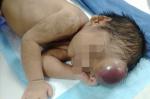
Our index case was delivered vaginally with an episiotomy at 38 weeks of gestation to a 28-year-old primi-gravida woman in Gynaecology Department of Datta Meghe Institute of Medical Sciences. During delivery the second stage was prolonged labor. At birth, there was a fluctuating swelling on frontal lobe that grew over time. Physical examination revealed an afebrile and well-appearing newborn with a huge, uniform, fluctuant swelling on the frontal area. The swelling is nothing but the accumulation of blood under the scalp. Therefore, a diagnosis of cephalohaematoma was determined based on the clinical characteristics. A cephalohematoma is characterised by the rupture of tiny blood vessels that cross the periosteum resulting in the accumulation of serosanguineous or bloody fluid between the periosteum and the skull. Small blood veins on the fetus's head are broken as a result of slight trauma during delivery. There is no test to identify cephalohematoma. The prominent protrusion on the newborn's head serves as the basis for diagnosis. A cephalohematoma is usually managed and treated through observation. Weeks pass before the cephalohematoma's bulk disappears as the clot-filled blood is gradually absorbed. The bulge may get firmer as the calcified blood collects over time. The reabsorption of the blood then begins. The patient was then instructed to visit for follow-up every eight days.
Figure 1: image of cephalohematoma
Search

This article authors
On Pubmed
On Google Scholar
Citation [Download]
Navigate this article
Similar articles in
Key words
Tables and figures
Article metrics
PlumX Metrics
A case of cephalohematomaRecently from the PAMJ-CM