Cytomegalovirus-induced petechial rash in an immunocompetent patient with acute hepatitis
Evangelia Christodoulou
Corresponding author: Evangelia Christodoulou, Department of Radiology, General University Hospital of Patras, Rion, Greece 
Received: 23 Jun 2026 - Accepted: 16 Aug 2026 - Published: 20 Aug 2026
Domain: Dermatology, Hepatology, Infectious disease, Internal medicine
Keywords: Petechial rash, exanthem, cytomegalovirus, infectious diseases, immunocompetency, hepatitis
Funding: This work received no specific grant from any funding agency in the public, commercial, or non-profit sectors.
©Evangelia Christodoulou et al. PAMJ Clinical Medicine (ISSN: 2707-2797). This is an Open Access article distributed under the terms of the Creative Commons Attribution International 4.0 License (https://creativecommons.org/licenses/by/4.0/), which permits unrestricted use, distribution, and reproduction in any medium, provided the original work is properly cited.
Cite this article: Evangelia Christodoulou et al. Cytomegalovirus-induced petechial rash in an immunocompetent patient with acute hepatitis. PAMJ Clinical Medicine. 2026;21:24. [doi: 10.11604/pamj-cm.2026.21.24.54168]
Available online at: https://www.clinical-medicine.panafrican-med-journal.com//content/article/21/24/full
Images in clinical medicine 

Cytomegalovirus-induced petechial rash in an immunocompetent patient with acute hepatitis
Cytomegalovirus-induced petechial rash in an immunocompetent patient with acute hepatitis
 Evangelia Christodoulou1,&
&Corresponding author

A 32-year-old woman presented with a 10-day history of rash and fatigue. Her medical history was significant for hypothyroidism treated with levothyroxine. Physical examination revealed erythematous punctate lesions on the lower extremities, discrete to confluent, without mucosal involvement, desquamation, lymphadenopathy, or other systemic signs. Laboratory evaluation showed a leukocyte count of 6.61×10³/μL with lymphocytic predominance (63.1%, absolute 4.17×10³/μL) and normal platelet count (193×103/μL). Inflammatory markers (ESR, CRP) and coagulation profile were within normal limits. Liver tests demonstrated marked hepatocellular injury with ALT 886 U/L and AST 566 U/L without cholestasis. Peripheral blood smear revealed atypical lymphocytes. Abdominal ultrasonography showed mild hepatosplenomegaly. Serology confirmed acute primary cytomegalovirus (CMV) infection (IgM positive, low-avidity IgG). Differential diagnosis included Epstein-Barr virus infection, autoimmune hepatitis, and drug-induced hypersensitivity reactions including DRESS syndrome. Infectious diseases specialists recommended conservative management, whereas hepatologists favored antiviral therapy given the severity of hepatitis; ultimately, a conservative approach was adopted with close monitoring. During follow-up, aminotransferases peaked at ALT 1149 U/L and AST 663 U/L, accompanied by nausea, right upper quadrant pain, and profound fatigue. The cutaneous eruption resolved within three weeks, and liver enzymes normalized within five weeks. At the six-week follow-up, serology showed increasing IgG avidity and declining IgM titers. In immunocompetent individuals, CMV infection is typically asymptomatic, and clinically significant disease remains underrecognized. Cutaneous involvement is rarely reported, and transaminase elevations rarely exceed fivefold the normal limit, as in this case. This case highlights an uncommon presentation of CMV infection in an immunocompetent adult with exanthem and severe acute hepatitis.
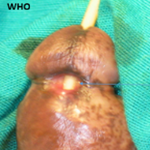
Figure 1: intraoperative view showing intact amniotic sac prolapsing through a lower uterine segment rupture along the prior caesarean scar
Search

This article authors
On Pubmed
On Google Scholar
Citation [Download]
Navigate this article
Similar articles in
Key words
Tables and figures

Article metrics
Recently from the PAMJ-CM