Postoperative discovery of a uterine sarcoma
Salah Houda, Amara Khouloud
Corresponding author: Salah Houda, Department of Gynecology and Obstetrics, Mohamed Tlatli Hospital, Nabel, Tunisia 
Received: 08 Jun 2026 - Accepted: 16 Aug 2026 - Published: 24 Aug 2026
Domain: Gynecology
Keywords: Fibroma, sarcoma, uterine
Funding: This work received no specific grant from any funding agency in the public, commercial, or non-profit sectors.
©Salah Houda et al. PAMJ Clinical Medicine (ISSN: 2707-2797). This is an Open Access article distributed under the terms of the Creative Commons Attribution International 4.0 License (https://creativecommons.org/licenses/by/4.0/), which permits unrestricted use, distribution, and reproduction in any medium, provided the original work is properly cited.
Cite this article: Salah Houda et al. Postoperative discovery of a uterine sarcoma. PAMJ Clinical Medicine. 2026;21:25. [doi: 10.11604/pamj-cm.2026.21.25.53840]
Available online at: https://www.clinical-medicine.panafrican-med-journal.com//content/article/21/25/full
Images in clinical medicine 

Postoperative discovery of a uterine sarcoma
Postoperative discovery of a uterine sarcoma
Salah Houda1,&, Amara Khouloud2
&Corresponding author

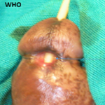
A 42-year-old female patient, with no significant past medical history and mother of three children born vaginally, presented to our emergency department with chronic pelvic pain that had been developing for several days without any other accompanying symptoms. On clinical examination, the patient was in good general condition and hemodynamically stable. Abdominal examination revealed a palpable, non-tender pelvic mass. Speculum examination showed a macroscopically normal cervix with signs of intrauterine bleeding. Breast examination was normal. An urgent beta-hCG test was negative. A transabdominal and transvaginal pelvic ultrasound showed an enlarged uterus containing a 10 cm intramural fibroid in the fundus, without any signs of atypia. The patient was discharged with analgesics and a pre-anesthetic consultation appointment for a possible hysterectomy. The patient returned for consultation after 3 days with heavy menorrhagia and severe anemia (hemoglobin = 5 g/dL). The decision was made to hospitalize the patient, administer intensive care, and receive a transfusion of isogroup and isorhesus packed red blood cells, followed by a laparoscopic hysterectomy. During the procedure, the surgeon noticed an unusual whitish appearance of the fibroid. He decided to perform an adnexectomy and await the results of the final histopathological examination of the surgical specimen. The histopathological examination confirmed a uterine sarcoma, and the case was discussed at a multidisciplinary team meeting. A thoracoabdominopelvic CT scan, ordered as part of the staging workup, came back normal. The patient underwent a second bilateral adnexectomy and received a chemotherapy protocol with a good outcome.
Figure 1: intraoperative findings of a uterine sarcoma
Search

This article authors
On Pubmed
On Google Scholar
Citation [Download]
Navigate this article
Similar articles in
Key words
Tables and figures

Article metrics
PlumX Metrics
Postoperative discovery of a uterine sarcomaRecently from the PAMJ-CM