Megaloblastic anemia in an infant revealing an inborn error of vitamin B12 metabolism: a case report
Kawtar Benkhaldoun, Wissal Nordine, Naima Fdil, Houda Nassih, Salma Rouhi, Wafa Quiddi, Sanae Sayagh
Corresponding author: Kawtar Benkhaldoun, Department of Hematology Laboratory, Mohammed VI University Hospital Center, Faculty of Medicine and Pharmacy of Marrakech, Cadi Ayyad University, Marrakech, Morocco 
Received: 07 May 2026 - Accepted: 28 May 2026 - Published: 11 Jun 2026
Domain: Laboratory medicine, Pediatric hematology
Keywords: Megaloblastic anemia, infant, functional vitamin B12 deficiency, methylmalonic aciduria, case report
Funding: This work received no specific grant from any funding agency in the public, commercial, or not-for-profit sectors.
©Kawtar Benkhaldoun et al. PAMJ Clinical Medicine (ISSN: 2707-2797). This is an Open Access article distributed under the terms of the Creative Commons Attribution International 4.0 License (https://creativecommons.org/licenses/by/4.0/), which permits unrestricted use, distribution, and reproduction in any medium, provided the original work is properly cited.
Cite this article: Kawtar Benkhaldoun et al. Megaloblastic anemia in an infant revealing an inborn error of vitamin B12 metabolism: a case report. PAMJ Clinical Medicine. 2026;21:8. [doi: 10.11604/pamj-cm.2026.21.8.53228]
Available online at: https://www.clinical-medicine.panafrican-med-journal.com//content/article/21/8/full
Case report 

Megaloblastic anemia in an infant revealing an inborn error of vitamin B12 metabolism: a case report
Megaloblastic anemia in an infant revealing an inborn error of vitamin B12 metabolism: a case report
 Kawtar Benkhaldoun1,&, Wissal Nordine1, Naima Fdil2, Houda Nassih3, Salma Rouhi1, Wafa Quiddi1,4, Sanae Sayagh1,2
&Corresponding author
Megaloblastic anemia in infants is a rare condition and may result from vitamin deficiencies or inborn errors of vitamin B12 metabolism. These disorders may present with severe hematological abnormalities, sometimes associated with neurological involvement, even when serum cobalamin and folate concentrations are normal, making the diagnosis challenging. We report the case of a four-month-old infant presenting with progressive pallor, severe pancytopenia, and early neurological abnormalities. Peripheral blood smear and bone marrow examination revealed dyserythropoiesis with megaloblastosis and signs of dysgranulopoiesis. Serum vitamin B12 and folate levels were normal; however, urinary organic acid analysis revealed isolated methylmalonic aciduria. The patient received intramuscular hydroxocobalamin. Clinical and hematological responses were rapid, with progressive normalization of the complete blood count and neurological improvement. This case illustrates the importance of metabolic investigations in megaloblastic anemia in infants, particularly in the presence of normal serum vitamin B12 levels. It highlights the diagnostic value of methylmalonic aciduria and the therapeutic benefit of hydroxocobalamin, even in cases of functional vitamin B12 deficiency.

Megaloblastic anemia in infants is a rare and potentially serious condition, most often reflecting impaired DNA synthesis in hematopoietic cells. Vitamin B12 and folate deficiencies represent the most common causes; however, less frequently described metabolic causes may also explain this morphological pattern and may involve defects in absorption, transport, or intracellular metabolism of cobalamin [1,2]. Functional vitamin B12 deficiencies may present with severe hematological abnormalities, sometimes associated with neurological involvement, even when serum vitamin B12 concentrations are normal [2]. This discrepancy between vitamin assay results and clinical manifestations should raise suspicion for an inborn error of vitamin B12 metabolism to ensure prompt and appropriate patient management. This work reports a case of an infant with cobalamin-responsive methylmalonic aciduria, identified during the investigation of megaloblastic anemia, and reviews the enzymatic disorders that may underlie these abnormalities.

Patient information: we describe the case of a four-month-old male infant admitted for progressive pallor and feeding difficulties. Pregnancy and delivery were unremarkable. There was no history of parental consanguinity. The family history was notable for the death of a sibling at the age of 10 months in the context of severe anemia of unknown etiology.
Clinical findings: clinical examination at admission revealed marked pallor and hypotonia, without tumor syndrome, organomegaly, or obvious signs of infection.
Timeline: the clinical course began at the age of two months with progressive mucocutaneous pallor, leading to hospital admission at four months of age. During hospitalization, the patient developed generalized tonic-clonic seizures, prompting further neurological evaluation. Initial laboratory investigations revealed pancytopenia, leading to further hematological assessment.
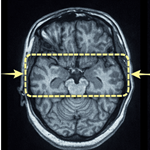
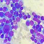
Diagnostic assessment: initial laboratory investigations showed pancytopenia, with hemoglobin at 7 g/dL, platelet count at 63 G/L, and leukocyte count at 3.9 G/L, including neutropenia at 0.4 G/L (Table 1). As part of bone marrow evaluation, a complete blood count performed in parallel with the bone marrow smear examination showed normochromic, normocytic, aregenerative anemia at 10.7 g/dL, thrombocytopenia at 41 G/L, and agranulocytosis at 0.38 G/L (Table 1). Bone marrow examination revealed a hypercellular marrow with numerous megakaryocytes showing hypolobulated nuclei. The erythroid lineage was quantitatively well represented (27%) and showed numerous signs of dyserythropoiesis, including megaloblastosis, nuclear-cytoplasmic asynchrony, stippled chromatin, binucleation, karyorrhexis, basophilic stippling, Howell-Jolly bodies, and cytoplasmic abnormalities. The granulocytic lineage was also well represented quantitatively (52%) and showed maturation abnormalities, with the presence of giant myelocytes and metamyelocytes, as well as hypersegmented neutrophils. No bone marrow infiltration by blast cells or cells of extra-hematopoietic origin was observed. No evidence of storage histiocytes or vacuolated erythroblasts was identified. All these findings were consistent with megaloblastic anemia (Figure 1). In the presence of marked megaloblastosis, vitamin assays were performed and revealed normal serum vitamin B12 levels at 231.9 pg/mL (reference range: 197-771) and normal folate levels at 16.9 ng/mL (reference range: 4.6-34.8). A bone marrow biopsy was performed but was non-contributory, yielding acellular material. Brain magnetic resonance imaging showed diffuse enlargement of the subarachnoid spaces suggestive of cerebral atrophy, associated with hypoplasia of the corpus callosum, without signal abnormalities in the white matter or basal ganglia. As part of the etiological investigation, a metabolic workup was performed. Plasma amino acid analysis by high-performance liquid chromatography, including phenylalanine, tyrosine, and leucine, was normal. Urinary screening for lysosomal storage diseases, including sulfatides, globosides (Gb3), hexosylceramides, lactosylceramides, sphingomyelins, phospholipids, and oligosaccharides, was negative. These results excluded classical aminoacidopathies and lysosomal storage disorders. However, urinary organic acid analysis revealed markedly increased excretion of methylmalonic acid (66-fold above normal), consistent with methylmalonic aciduria. Additional metabolic investigations, including plasma homocysteine measurement and genetic testing, could not be performed during the patient´s evaluation.
Diagnosis: based on the association of megaloblastic anemia, neurological manifestations, isolated methylmalonic aciduria, and normal serum vitamin B12 levels, a cobalamin-related metabolic disorder causing functional vitamin B12 deficiency was strongly suspected.
Therapeutic intervention: the patient received intramuscular hydroxocobalamin at a dose of 1mg according to the following regimen: one daily injection for three consecutive days during the first week, followed by one daily injection for two days during the second week, and then one weekly injection thereafter. Prolonged treatment was planned considering the suspected underlying metabolic vitamin B12 disorder. Given the severity of the anemia, the patient also received several packed red blood cell transfusions.
Follow-up and outcome: the clinical course was marked by rapid clinical and hematological improvement following initiation of treatment. Progressive normalization of complete blood count parameters was observed during follow-up, with improvement of hemoglobin level (13.1 g/dL), leukocyte count (14.1 G/L), and platelet count (526 G/L) (Table 1).
Patient perspective: as the patient was an infant, the parents reported noticeable clinical improvement after treatment and expressed adherence to the proposed follow-up plan. They were grateful for the care and support provided by the medical and nursing staff to their infant.
Informed consent: the informed consent for publication of this case report was obtained from the patient´s parents.
Megaloblastic anemia is a rare entity in pediatrics [2,3], with an estimated incidence of approximately 0.014% [3]. It is most commonly caused by vitamin B12 or folate deficiency [4], whereas congenital causes, particularly inborn errors of cobalamin metabolism, are uncommon and have been reported in only a limited number of cases in the literature [1]. In this infant, the association of megaloblastic anemia, early neurological manifestations, and the presence of methylmalonic aciduria despite normal serum vitamin B12 levels suggest a defect in intracellular cobalamin metabolism. Indeed, plasma vitamin B12 levels may remain normal in several inherited errors of cobalamin metabolism, particularly when the defect involves its transport or intracellular utilization rather than its intestinal absorption [1,2]. The marked clinical and biological improvement following the initiation of hydroxocobalamin therapy strongly supports the diagnosis of functional cobalamin deficiency, even in the presence of initially normal serum levels [5]. Notably, serum cobalamin measurement does not reliably reflect its intracellular utilization or the activity of vitamin B12-dependent metabolic pathways. Therefore, functional markers such as methylmalonic acid or homocysteine are of particular interest for confirming a metabolic vitamin B12 deficiency [5,6]. This favorable response to treatment has been classically described both in transcobalamin II deficiency [7] and in certain inborn errors of cobalamin metabolism [1]. Several pediatric cases report early presentation with severe hematological involvement and rapid improvement under parenteral hydroxocobalamin, as observed in our patient [1,8].
Inborn errors of cobalamin metabolism involve defects affecting the intracellular transport, processing, or utilization of vitamin B12, resulting in distinct biochemical and clinical phenotypes [1,2]. Depending on the affected metabolic pathway, these disorders may lead to isolated methylmalonic aciduria, hyperhomocysteinemia, or combined metabolic abnormalities, often associated with hematological and neurological manifestations of variable severity [1,8]. This case highlights the diagnostic challenge when comprehensive metabolic and genetic investigations are not fully accessible in a given clinical context, as plasma homocysteine levels and genetic confirmation were not performed, limiting precise classification of the underlying defect. The presence of isolated methylmalonic aciduria may suggest involvement of the adenosylcobalamin-dependent pathway; however, combined cobalamin metabolic defects could not be excluded due to the incomplete metabolic and genetic evaluation [9]. Nevertheless, the association of megaloblastic anemia, methylmalonic aciduria, neurological manifestations, and favorable response to hydroxocobalamin strongly supported the diagnosis of functional vitamin B12 deficiency.
Although not specific in a four-month-old infant, diffuse enlargement of the subarachnoid spaces suggestive of cerebral atrophy, associated with hypoplasia of the corpus callosum, should be considered as possible neurological complications of vitamin B12 deficiency or its metabolic pathways. These findings are consistent with the spectrum of neurological manifestations described in metabolic cobalamin disorders. Neurological involvement in these disorders is well documented and may occur early in infancy, even in the absence of overt vitamin deficiency. The accumulation of methylmalonic acid and impaired methionine synthesis are thought to play a central role in neurotoxicity, leading to delayed myelination, cerebral atrophy, and neurodevelopmental abnormalities. These mechanisms are supported by previous studies demonstrating that both methylmalonic acid accumulation and reduced methionine availability, with subsequent disruption of methylation processes, contribute to neuronal dysfunction and abnormal brain development [10]. Early diagnosis and timely management are crucial to minimize long-term neurological sequelae.
The present case illustrates the diagnostic complexity of megaloblastic anemia in infants. It highlights that normal vitamin assay results do not exclude functional cobalamin deficiency and emphasizes the value of an integrated approach combining bone marrow morphological assessment and metabolic investigations. Early identification and management with parenteral hydroxocobalamin allow rapid correction of hematological abnormalities and help prevent neurological complications. This case also underlines the need for a multidisciplinary approach and access to metabolic and genetic investigations to ensure accurate diagnosis and optimal management.
The authors declare no competing interests.
All authors contributed to the management of the patient. They have read and approved the final version of this manuscript.
Table 1: complete blood count parameters before and after hydroxocobalamin therapy
Figure 1: peripheral blood smear and bone marrow examination showing morphological abnormalities consistent with megaloblastic anemia: A) peripheral blood smear showing hypersegmented neutrophils; B) bone marrow smear at low magnification showing increased cellularity; C, D) bone marrow at high magnification showing megaloblastic changes
- Watkins D, Rosenblatt DS. Inborn errors of cobalamin absorption and metabolism. Am J Med Genet C Semin Med Genet. 2011;157(1):33-44. PubMed | Google Scholar
- Rosenblatt DS, Whitehead VM. Cobalamin and folate deficiency: acquired and hereditary disorders in children. Semin Hematol. 1999;36(1):19-34. PubMed | Google Scholar
- Belhaj R, Maaloul I, Kolsi R, Rekik T, Chabchoub I, Aloulou H et al. Study of clinical manifestations and etiologies of megaloblastic anemia in children. Transfus Clin Biol. 2025;32(2):159-163. PubMed | Google Scholar
- Green R, Mitra AD. Megaloblastic anemias: nutritional and other causes. Med Clin North Am. 2017;101(2):297-317. PubMed | Google Scholar
- Hunt A, Harrington D, Robinson S. Vitamin B12 deficiency. BMJ. 2014;349:g5226. PubMed | Google Scholar
- Monsen ALB, Ueland PM. Homocysteine and methylmalonic acid in diagnosis and risk assessment from infancy to adolescence. Am J Clin Nutr. 2003;78(1):7-21. PubMed | Google Scholar
- Ünal S, Karahan F, Arkoğlu T, Akar A, Kuyucu S. Different presentations of patients with transcobalamin II deficiency: a single-center experience from Turkey. Turk J Hematol. 2019;36(1):37-42. PubMed | Google Scholar
- Carrillo-Carrasco N, Chandler RJ, Venditti CP. Combined methylmalonic acidemia and homocystinuria, cblC type. I. Clinical presentations, diagnosis and management. J Inherit Metab Dis. 2012;35(1):91-102. PubMed | Google Scholar
- Watkins D, Rosenblatt DS. Inherited defects of cobalamin metabolism. Vitam Horm. 2022;119:355-376. PubMed | Google Scholar
- Du M, Li M, Wu S, Wu X, Chen Y, Zhu C. Metabolic toxicity and neurological dysfunction in methylmalonic acidemia: from mechanisms to therapeutics. Mol Med. 2025;31(1):333. PubMed | Google Scholar
Search

This article authors
On Pubmed
On Google Scholar
Citation [Download]
Navigate this article
Similar articles in
Key words
Tables and figures


Article metrics
Recently from the PAMJ-CM