Clinical presentation and surgical outcomes of penile fracture in Tamale Teaching Hospital: a three-year case series
Benjamin Akinkang, Bentil Awe Wewoli, Justine Dakurah
Corresponding author: Benjamin Akinkang, Department of Pathology, University for Development Studies, Tamale, Ghana 
Received: 30 Apr 2026 - Accepted: 28 May 2026 - Published: 11 Jun 2026
Domain: Health emergencies, General surgery, Urology
Keywords: Penile fracture, popping sound, Northern Ghana, eggplant deformity, corpus carvenosum, case series
Funding: This research received no specific grant from any funding agency in the public, commercial, or not-for-profit sectors.
©Benjamin Akinkang et al. PAMJ Clinical Medicine (ISSN: 2707-2797). This is an Open Access article distributed under the terms of the Creative Commons Attribution International 4.0 License (https://creativecommons.org/licenses/by/4.0/), which permits unrestricted use, distribution, and reproduction in any medium, provided the original work is properly cited.
Cite this article: Benjamin Akinkang et al. Clinical presentation and surgical outcomes of penile fracture in Tamale Teaching Hospital: a three-year case series. PAMJ Clinical Medicine. 2026;21:9. [doi: 10.11604/pamj-cm.2026.21.9.53087]
Available online at: https://www.clinical-medicine.panafrican-med-journal.com//content/article/21/9/full
Case series 

Clinical presentation and surgical outcomes of penile fracture in Tamale Teaching Hospital: a three-year case series
Clinical presentation and surgical outcomes of penile fracture in Tamale Teaching Hospital: a three-year case series
 Benjamin Akinkang1,&, Bentil Awe Wewoli2, Justine Dakurah2
&Corresponding author
Penile fracture is a rare urological emergency caused by rupture of the tunica albuginea of the corpora cavernosa, typically in the erect state. Despite its characteristic presentation, data from low-resource settings remain limited. This study describes the presentation, diagnosis, and outcomes of penile fracture in Northern Ghana. We conducted a retrospective case series of patients managed for penile fracture at Tamale Teaching Hospital between January 2023 and December 2025. Data on demographics, mechanism of injury, time to presentation, clinical findings, diagnostic approach, operative details, and outcomes were analysed using descriptive statistics. Nine patients were managed during the study period, with a mean age of 33.89 years (range: 22-47 years). The most common causes of injury were manipulation of the erect penis (44.4%), coitus (33.3%), and falls onto an erect penis (22.2%). The mean time to presentation was 1.72 hours. All patients presented with the classical triad of a “popping” sound, pain, and rapid detumescence, with “eggplant” deformity observed in all cases. Diagnosis was primarily clinical, with ultrasonography required in one case. Surgical repair was performed in all patients via a subcoronal degloving approach. Urethral injury was identified in 33.3% of cases. The mean hospital stay was 3 days. At the 3-month follow-up, all patients had satisfactory erectile function with no reported complications. Penile fracture in this setting demonstrates a consistent clinical presentation and excellent outcomes following prompt surgical intervention. Early presentation and timely repair are key to preventing complications, even in resource-limited environments.

Penile fracture is a relatively rare but well-recognized urological emergency characterised by traumatic rupture of the tunica albuginea of the corpora cavernosa, typically occurring when the penis is in an erect state [1]. It is most commonly associated with blunt trauma during sexual intercourse, although other mechanisms such as masturbation, rolling over in bed, or direct trauma have been described [2]. Despite its dramatic presentation and potential for significant morbidity, the true incidence of penile fracture is difficult to ascertain, particularly in low- and middle-income settings, where sociocultural factors, stigma, and underreporting [3] contribute to the limited epidemiological data [4]. Clinically, penile fracture often presents with a characteristic triad of an audible “cracking” or “popping” sound, immediate penile pain, rapid detumescence, and subsequent swelling leading to the classical “eggplant deformity.” [5]. Diagnosis is largely clinical, based on history and physical examination, with imaging modalities such as ultrasonography or magnetic resonance imaging reserved for atypical cases or when associated urethral injury is suspected [5]. Early recognition is crucial, as delayed diagnosis and management are associated with complications, including erectile dysfunction, penile curvature, painful intercourse, and urethral strictures [6].
Management of penile fracture has evolved, with immediate surgical repair now widely regarded as the gold standard of treatment [7]. Surgical exploration allows evacuation of haematoma and repair of the tunical tear, significantly reducing long-term complications compared to conservative approaches [7]. Outcomes following prompt surgical intervention are generally favourable, with high rates of erectile function preservation and minimal morbidity reported across multiple studies [8]. In Ghana, penile fracture remains an uncommon but increasingly reported condition, largely documented through case reports and small case series [9,10,11]. These findings highlight both the consistency of clinical presentation and the challenges related to delayed presentation in the local context. Despite these emerging reports, there remains a paucity of comprehensive data on the presentation patterns, diagnostic approaches, and outcomes of management of penile fracture in Northern Ghana. Variations in health-seeking behaviour, access to specialist urological care, and resource availability may influence both the timing of presentation and treatment outcomes. Consequently, there is a need for more robust local data to better characterise this condition within the Ghanaian setting. This case series, therefore, aims to describe our experience with the presentation, diagnosis, and management of penile fracture in Northern Ghana, with particular emphasis on clinical patterns, treatment modalities, and outcomes. Such data are essential to inform clinical practice, improve early recognition, and optimise management strategies in resource-limited settings.

This case series was conducted at the Urology Unit of the Department of Surgery at the Tamale Teaching Hospital, a referral facility for the Northern parts of Ghana. Ethical clearance was waived, and written consent was taken from the patients. We retrospectively reviewed all cases of penile fractures managed in the department from January 2023 to December 2025. Information on age, time between onset and presentation to the health facility, and cause of injury. Other information includes: means of diagnosis (clinical or radiological), physical appearance of the penile shaft, site of injury, urethral involvement, the number of days on admission, and the maintenance of good erection upon review. This information is summarised as patient information and clinical presentation in Table 1. All the 9 patients presented with the history of hearing a popping sound, rapid detumescence, and pain within the penis. Figure 1 is a fractured penis presenting with the characteristic “eggplant” deformity.
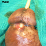
Operative techniques: all the patients had surgical intervention. Under spinal anaesthesia, the patients were prepped and draped. Under aseptic technique, a subcoronal degloving incision was made. The fracture sites, which were transverse tears located at the proximal penile shaft, were exposed (Figure 2), as shown in Table 2. For cases with urethral injury as shown in Figure 2, a 16 French urethral catheter was passed, and the urethral ruptures were repaired via simple primary closure with Vicryl 3/0. All the tunica albuginea lacerations were repaired with nylon 3/0. The wounds were closed with Vicryl 0 or Vicryl 2/0. The postoperative condition of the patients was satisfactory, and the patients were discharged from the hospital. Patients with urethral involvement went home with a catheter in situ. The catheters were subsequently removed after 4 weeks post-op. Voiding was satisfactory without lower urinary tract obstruction. All patients were reviewed 3 months after discharge, and all had good erections. No other complications were noted. Data was entered into and analysed using Microsoft Excel. Simple descriptive statistics, such as proportions, mean, and range, were used.
The hospital saw and managed 9 cases of penile fracture from 2023 to 2025. Three (3) out of the 9 (33.3%) were referred, while the remaining 6 reported directly to the accident and emergency unit in a teaching hospital (TTH). The mean age of presentation was 33.89 (SD, 8.31) with a range of 22 to 47 years. The mean time between injury and presentation to the hospital was 1.72 (SD, 1.66) with a range of 0.5 to 5 hours. Four out of the 9 (44.4%) reported manipulation of the erect penis as the cause of injury, 3 (33.3%) reported coitus as the cause, and the remaining 2 were as a result of falling on an erect penis. All the cases show “eggplant” deformity. Aside from one case that required an ultrasound for diagnosis, the rest were diagnosed clinically. Only one case involved both bilateral corpora cavernosa. Three (33.3%) patients had right corpus cavernosum rupture, 2 had left corpus cavernosum, and 3 also had ventral rupture. The 3 with the ventral rupture also had urethral involvement. The widest dimension of the defects ranged from 1 to 4 cm, with a mean dimension of 2.11 cm (SD, 0.82). All the injuries were located at the proximal penile shaft. The number of days on admission ranges from 2 to 5, with a median of 3 days. There was no postoperative complication. All patients had good erections after 3 months of follow-up review.
Penile fracture is a rare urological emergency that is becoming increasingly common, especially in our jurisdiction. The rarity is due to the mobility of the genital area, making the penis less vulnerable to trauma and subsequent fracture [12]. The true prevalence cannot be accurately ascertained due to underreporting [10]. The reasons for underreporting may be ascribed to patients feeling embarrassed due to the sexually related causes [13]. The fracture involves disruption of the tunica albuginea of the corpus cavernosa [5]. Occasionally, the corpus spongiosum and the urethra may be involved [5]. Penile fracture occurs over a wide age range. A series by Molla et al. involving 18 patients also estimated the mean age at 37, with an age range of 24 to 70 years [14]. The ages of the participants in this series range from 22 to 47, with a mean age of 33.89. In a systematic review of 438 patients by Falcone et al. the mean age was 36 [8], which is not much different from the 33.89 in our study. The causes of penile fracture, among others, include: blunt trauma to the erect penis during sexual intercourse, falling on the erect penis, masturbation, forcefully bending the erect penis, and manipulating the erect penis to achieve detumescence [2]. In our series, the causes in a decreasing frequency include: manipulating the erect penis to achieve detumescence, coitus or sexual intercourse and falling on the erect penis. Sexual intercourse as the cause of injury appears to lead in many of the series [5,11].
When the fracture occurs, the patient will usually hear a cracking or popping sound, followed by rapid detumescence and pain. Swelling will later occur, causing the shaft to deviate to give the characteristic “eggplant” picture [5]. All our 9 cases reported the popping sound, the rapid detumescence, and the pain. In the series by Molla et al. all 18 patients had penile pain and swelling, a penile distortion was present in 15 people (83%), and 13 patients (72%) reported hearing a cracking (popping) sound that causes penile swelling, pain, and fast detumescence [14]. Time is of utmost importance if long term complication from the penile fracture is to be avoided [6]. Many factors, such as socioeconomic factors, financial constraints, embarrassment, etc., may cause a delay in reporting to the hospital [3]. In our series, the mean time duration was 1.72 hours, with some reporting as early as 0.5 hours and others as late as 5 hours. Some authors have reported a time range of 7 hours to 1 month [14] and 3 hours to 10 days [11]. It can be clearly seen that all the cases in this series were reported early to the facility, despite the fact that some were referred from other peripheral facilities.
The diagnosis of all except one case was straightforward based on history and clinical findings. Only one case needed the help of ultrasonography to confirm. This is in line with existing literature [5]. A case series from Cape Coast, Ghana, demonstrated that diagnosis was made clinically in all patients [10]. In their series of 18 patients, Molla et al. used only clinical examination to diagnose 11 patients and an ultrasound scan to confirm the remaining 7 patients [14]. Falcone et al. also revealed in their systematic review that almost all authors employed radiological scans to estimate the extent of penile fracture, but not for diagnosis [8]. This goes to reemphasise the critical role history and physical examination play in the diagnosis of penile fracture, especially in resource-limited settings where access to radiological services may be limited. The fratures are commonly located in the penile shaft and more ventrally [5,14]. Ofori et al. reported 70.6% of their fracture to have occurred in the ventral side [11]. The tunica layers are thinner on the ventral side than the dorsal side, contributing to the high risk of fractures in that area and the vulnerability of the urethra in a fracture [8]. The more ventral urethra is prone to damage [11]. As seen in 3 of these cases, all the ventrally located fractures had associated urethral injury.
The sizes of defects in this series range from 1 to 4 cm, with a mean size of 2.11 cm in the widest dimension. This is not so different from the findings of Ofori et al. who reported 0.5 to 3 cm [11]. Molla et al. also reported similar sizes [14]. The distribution of our fracture sites is in line with the literature [5,8]. In this case series, 33.3% of the fractures occurred at the right corpus cavernosum, and 33.3% at the ventral side, which is in line with Ofori et al. [11]. Immediate surgical intervention is required to achieve good results and to reduce hospital stay [15]. While most recent authors advocate for immediate surgical repair for fractures with or without urethral involvement [5,7], conservative management can sometimes be employed if there is delayed presentation or in special cases [14]. Early surgical intervention is noted to be associated with fewer complications compared to conservative management [5]. It also results in better outcomes in terms of reduced penile curvature, erectile dysfunction, etc. [5]. Ofori et al. performed immediate surgery for 70.6% of their 17 cases [11]. Molla et al. performed surgery for 77.7% of their 18 cases, with only 4 managed conservatively due to a delay of more than a week before presentation [14]. All 9 cases in this series had immediate surgery with subsequent good outcomes.
A circumferential incision was made just beneath the corona, and the skin was degloved to expose the injury sites. This kind of approach, which is the most common approach, gives the surgeon a better vision, helping locate the injury site [8]. The choice of suture type to close the tunica defect depends on the surgeon and the type available. Proponents of employing non-absorbable sutures to close tunical defects contend that they give the tunical edges long-lasting support, assisting in maintaining their alignment and preventing the defect's breakdown or recurrence during times of elevated intracorporeal pressure [7]. In this case series, the urethral injuries were sutured using Vicryl in an interrupted manner. The corpus cavernosa and tunica were closed using nylon, also in an interrupted fashion. Postoperative review after 3 months showed full recovery, restoration of good erection and no other complications. Ofori et al. reported 17.6% of their patient had wound infection, 23.3% hard short-term complications such as penile curvature and painful sex, and 41. 2% had erectile dysfunction [11]. Even though the huge sample size of Ofori et al. series may account for this, the increased mean duration of injury before reporting may have also contributed to these complications [6,11].
Limitation: this study has several limitations. The small sample size and single-centre design limit the generalisability of the findings to the broader population. The relatively short follow-up period of 3 months may not adequately assess long-term complications such as erectile dysfunction, penile curvature, or urethral stricture. Additionally, the absence of a comparison group precludes evaluation of outcomes between different management approaches.
Penile fracture, though uncommon, is a true urological emergency with a characteristic clinical presentation that allows for prompt diagnosis even in resource-limited settings. This case series from Northern Ghana demonstrates that most patients present early and can be accurately diagnosed based on history and physical examination without reliance on advanced imaging. Immediate surgical exploration and repair using a subcoronal degloving approach resulted in excellent outcomes, including preservation of erectile function and absence of postoperative complications at short-term follow-up. Our findings reinforce the growing body of evidence that early surgical intervention is the gold standard for the management of penile fracture, including cases with associated urethral injury. The uniformly favourable outcomes observed in this series highlight the importance of timely presentation and access to surgical care. Given the potential for underreporting due to sociocultural stigma, there is a need for increased public awareness to encourage early health-seeking behaviour. Additionally, strengthening urological services and surgical capacity in peripheral centres may further improve outcomes. Larger, multicentre studies with longer follow-up are recommended to better characterise long-term complications and functional outcomes in this setting.
What is known about this topic
- Penile fracture is increasingly becoming common in Ghana;
- There is an underreporting of penile fracture due to embarrassment and socioeconomic reasons;
- Rapid surgical correction leads to better outcomes with minimal complications.
What this study adds
- This study reemphasises the growing body of evidence that early surgical intervention is the gold standard for the management of penile fracture;
- The uniformly favourable outcomes observed in this series highlight the importance of timely presentation and access to surgical care.
The authors declare no competing interests.
All authors were involved in the conceptualisation, analysis, and writing up of this work. All the authors have read and approved the final version of this manuscript.
Table 1: information and clinical presentation of patients managed for penile fracture in TTH from January 2023 to December, 2025
Table 2: operative and postoperative findings of patients managed for penile fracture in TTH from January 2023 to December, 2025
Figure 1: fractured penis with “eggplant” deformity of one of the patients managed for penile fracture in TTH from January 2023 to December, 2025
Figure 2: defects in the tunica and corpus cavernosum in the proximal penile shaft of two of the patients managed for penile fracture in TTH from January 2023 to December, 2025
- Efendi A W. Biomechanical Analysis of Penile Erection: Comparative Study Using LISA FEA. MERJMB. April, 2025;5(2):26-33. Google Scholar
- Fredrick A, Vanni AJ. Traumatic penile injuries. A Clinical Guide to Urologic Emergencies. 2021 Aug 6:179-88. Google Scholar
- Eke N. Fracture of the penis. Br J Surg. 2002 May;89(5):555-65. PubMed | Google Scholar
- Imran M, Kamran A, Tanveer A, Farho MA. Penile fracture: A case report. Int J Surg Case Rep. 2023 Sep;110:108749. PubMed | Google Scholar
- Amer T, Wilson R, Chlosta P, AlBuheissi S, Qazi H, Fraser M et al. Penile fracture: A meta-analysis. Urol Int. April 2016;96(3):315-329. PubMed | Google Scholar
- El-Assmy A, El-Tholoth HS, Mohsen T, Ibrahiem el HI. Long-term outcome of surgical treatment of penile fracture complicated by urethral rupture. J Sex Med. 2010 Nov;7(11):3784-8. PubMed | Google Scholar
- Katswere J, Bahloul A, Hounsou R, Todalehou S, Kaduku G, Byabene D et al. Diagnostic and management of penile fracture: a literature review. J Med Res. 2019;5(4):162-4. Google Scholar
- Falcone M, Garaffa G, Castiglione F, Ralph DJ. Current Management of Penile Fracture: An Up-to-Date Systematic Review. Sex Med Rev. April 2018;6(2):253-260. PubMed | Google Scholar
- Dakurah J, Akinkang B, Yenli EMT, Awe BA, Musah Z. Penile Fracture, Unusual Mechanism of Injury: A Case Report. Asian J Adv Res Rep. July 2025;19(7):101-106. Google Scholar
- Assabill F, Ofori EO, Maison PO, Asante-Asamani A. Penile fracture–Report on three cases from Cape Coast, Ghana. Open J Urol. 2022;12:146-56. Google Scholar
- Ofori EO, Adanu KK, Akpakli EA, Mensah JE. Penile Fracture: Our Experience in Korle Bu Teaching Hospital in Accra, Ghana. J West Afr Coll Surg. 2025 Oct-Dec;15(4):400-406. PubMed | Google Scholar
- Tolani M A, Webber R, Buckley L. Penile trauma burden and aetiology in the paediatric and adult population: A scoping review and critical analysis of the literature. J West Afr Coll Surg. 2024 Jan-Mar;14(1):5-16. PubMed | Google Scholar
- Wani I. Management of penile fracture. Oman Med J. 2008 Jul;23(3):162-5. PubMed | Google Scholar
- Molla YD, Erku BA, Kassa SA, Alemu HT, Mekonnen DC. Penile fracture: a case series of 18 patients. Ann Med Surg (Lond). 2023 Oct 12;85(12):6186-6191. Google Scholar
- Baba D, Ekici N, Taşkiran AT, Şenoğlu Y, Başaran E, Balik AY. Complete urethral rupture accompanying penile fracture: a case report. BMC Urol. Dec, 2025;25(1):1-6. PubMed | Google Scholar
Search

This article authors
On Pubmed
On Google Scholar
Citation [Download]
Navigate this article
Similar articles in
Key words
Tables and figures



Article metrics
Recently from the PAMJ-CM