Airway management of a patient with post-burn scar contractures on the face and neck: a case report
Megan Males, Blaise Bayingana
Corresponding author: Megan Males, University of Witwatersrand, Faculty of Health Sciences, Department of Anaesthesiology, Johannesburg, South Africa 
Received: 25 Mar 2026 - Accepted: 28 May 2026 - Published: 12 Jun 2026
Domain: Emergency medicine,Intensive care medicine,Anesthesiology
Keywords: Airway management, post-burn scar contractures, mask ventilation, video-assisted laryngoscopy, case report
Funding: This work received no specific grant from any funding agency in the public, commercial, or not-for-profit sectors.
©Megan Males et al. PAMJ Clinical Medicine (ISSN: 2707-2797). This is an Open Access article distributed under the terms of the Creative Commons Attribution International 4.0 License (https://creativecommons.org/licenses/by/4.0/), which permits unrestricted use, distribution, and reproduction in any medium, provided the original work is properly cited.
Cite this article: Megan Males et al. Airway management of a patient with post-burn scar contractures on the face and neck: a case report. PAMJ Clinical Medicine. 2026;21:11. [doi: 10.11604/pamj-cm.2026.21.11.52377]
Available online at: https://www.clinical-medicine.panafrican-med-journal.com//content/article/21/11/full
Case report 

Airway management of a patient with post-burn scar contractures on the face and neck: a case report
Airway management of a patient with post-burn scar contractures on the face and neck: a case report
![]() Megan Males1,2,&,
Megan Males1,2,&, ![]() Blaise Bayingana1,3
Blaise Bayingana1,3
&Corresponding author
Securing an airway is crucial when providing anaesthesia for patients with burn injuries, due to the high likelihood of a difficult airway. This case report describes a 35-year-old male with electrical burns, who presented for elective left upper eyelid reconstruction. Airway management was challenging due to post-burn scar contractures and facial scarring. Patient declined awake fibre-optic intubation; the anaesthetic plan, therefore, included inhalational induction with spontaneous breathing, video-laryngoscope-guided intubation, multimodal analgesia, and awake extubation. Burn injuries pose a significant public health concern in low- and middle-income countries like South Africa. Burn patients present complex anaesthetic challenges, including difficult airway management, fluid resuscitation, the need for specialised monitoring, and thermoregulation. When managing a difficult airway, awake fibre-optic intubation is considered the gold standard. However, alternative techniques, such as inhalational induction, supraglottic airway devices, and invasive techniques, may be used. In conclusion, thorough preparation and adherence to best practice guidelines will enable successful airway management.

When providing anaesthesia to patients with burn injuries, securing an airway is crucial as these patients often have difficult airways. Challenges involving mask ventilation, laryngoscopy, intubation, and/or cricothyroidotomy should be appropriately prepared for. Likewise, great care should be taken to avoid a 'cannot intubate, cannot ventilate' situation, as mortality and morbidity in these patients is directly related to the inability to deliver oxygen. This case report discusses the airway management of a patient with post-burn scar contractures on the face and neck, presenting for reconstructive surgery.

Patient information: a 35-year-old male presented for elective left upper eyelid reconstruction with a skin graft. On history, it was noted that he sustained 40% electrical burns to his face, neck, and left upper limb two months prior. He was haemodynamically unstable requiring intubation, resuscitation with inotropes, and admission to the intensive care unit (ICU). During this initial admission, he underwent multiple burn wound debridements and a left upper limb amputation. After one month of inpatient care, he was discharged home. On further history, he is a smoker with an eight pack-year history, has no reported comorbidities, and maintains good functional capacity. He now presents for elective ophthalmologic surgery as part of his reconstructive journey.
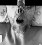
Clinical findings: preoperatively, the patient´s vital signs were within normal limits, and he had a normal respiratory and cardiovascular examination. He was assessed as having anticipated difficult mask ventilation, laryngoscopy, and intubation. However, palpation of the neck confirmed an identifiable cricothyroid membrane, should a rescue airway have been required. The patient had extensive burn scars covering his entire face and neck, with a tip amputation of his nose and scarred, narrow nasal passages. Multiple post-burn contractures restricted both neck extension and flexion and limited mouth opening to one and a half fingers (Figure 1). He had poor dentition and a class IV Mallampati score. Difficult intravenous access was anticipated due to left upper arm amputation and right arm burn scars, so central venous access was considered as an alternative. The patient was graded as an American Society of Anesthesiologists (ASA) I.
Timeline: not applicable, as this case report focuses on a single acute perioperative episode. A chronological account is detailed in the 'Intervention' section.
Diagnostic assessment: not applicable, as this case report focuses on anaesthesia management rather than diagnostic assessment and diagnosis.
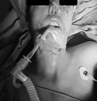
Intervention: given the anticipated difficult airway, the patient was counselled on awake fibre-optic intubation. However, due to heightened preoperative anxiety, the patient declined this option. Consequently, the anaesthetic plan involved a general anaesthetic with inhalational induction to maintain spontaneous breathing, video-laryngoscope-assisted intubation, and the presence of a senior anaesthetist in the theatre. Standard ASA monitors (pulse oximetry, non-invasive blood pressure monitor, and continuous electrocardiogram) were applied. 18G intravenous access was successfully achieved in the right forearm. An inhalational induction with sevoflurane, air, and oxygen was then done. Mask ventilation was possible, and video laryngoscopy was used to visualise the vocal cords. Once the vocal cords were visualised, propofol (1 mg/kg) and fentanyl (2 mcg/kg) were administered intravenously. A size 7.0 endotracheal tube (ETT) was selected and placed using an introducer. Placement was confirmed via capnography and auscultation, and the tube was secured (Figure 2). A oesophageal temperature probe was inserted, and the patient was covered with a Bair Hugger (forced air warming system). Muscle relaxants were avoided to maintain spontaneous breathing and minimise the risk of a 'cannot intubate, cannot ventilate' scenario. Intraoperatively, anaesthesia was maintained with sevoflurane, air, and oxygen, with pressure support ventilation. The patient received multi-modal analgesia (intravenous paracetamol 1 g and morphine 0.5 mg/kg) and dexamethasone (4 mg) to reduce airway oedema.
Follow-up and outcome: the patient was extubated awake and transferred to the recovery room with normal haemodynamic parameters and an oxygen saturation of 98% on room air. Following an uncomplicated recovery room stay, the patient was discharged to the ward for routine postoperative care.
Patient perspective: a formal patient perspective was not obtained as the patient was under general anaesthesia during the management described. However, having declined awake fibre-optic intubation due to preoperative anxiety, the patient subsequently expressed satisfaction with the alternative induction technique.
Informed consent: the patient provided written informed consent for the publication of their medical information and accompanying photographs. Identifying details have been removed to maintain patient anonymity and confidentiality.
Burns are devastating injuries and pose a significant public health concern. Globally, there are approximately 8 000 000 new burn cases each year, with over 95% of burn deaths occurring in low- and middle-income countries [1]. Rode et al. [2] estimate that about 3.2% of South Africa´s population will suffer from burn injuries each year. A South African survey revealed that burns are the most common external cause of death in children younger than four and the third most common cause in children younger than 18 [2]. Electrical burns, in particular, are an increasing concern in South Africa, accounting for 6% of all burn injuries [2]. This is largely attributed to illegal electrical connections, exposed wires, and cable theft [2]. Furthermore, limited access to electricity leading to widespread paraffin use, combined with socioeconomic factors such as overcrowding, poverty, and urban migration contribute to the high incidence of burns in South Africa [2].
Burn injuries can be classified as major or minor [1]. Major burns require intervention from multiple specialist services, while minor burns can be effectively managed by nurse practitioners and general medical doctors [2]. A burn is classified as major if it involves partial thickness burns (involving the epidermis and dermis) affecting >10% total body surface area, any full-thickness burns (involving the epidermis, dermis, and subcutaneous fat), burns involving the face, hands, feet, genitalia, or major joints, as well as chemical burns, electrical burns, lightning strike injuries, significant inhalation injuries, or burns in patients with multiple medical disorders or associated traumatic injuries [3]. Notably, 14% of burn injuries in South Africa are classified as major and therefore require intervention from multiple specialist services [2].
Anaesthesia providers are vital in the perioperative care of burn patients, providing airway management, intraoperative anaesthesia, and postoperative analgesia [3]. Burn injury management is typically divided into two phases: i) the acute phase involves wound debridements and decompressive procedures; ii) the reconstructive phase may involve skin grafting, tissue expansion, or free flap transfers [1].
Burn patients pose unique anaesthetic challenges due to their complex injuries and resulting physiological changes [1,4]. Airway management is a critical consideration as burn patients are frequently classified as having a difficult airway [1,4]. The ASA defines a difficult airway as a situation where challenges are encountered in facemask ventilation, laryngoscopy, ventilation using a supraglottic airway, intubation, extubation, or invasive airway placement [5]. According to Bittner et al. [4], facial swelling, painful wounds with slippery exudate, dressings, and nasogastric tubes can complicate facemask ventilation. Furthermore, thermal damage resulting in glottic swelling, along with restricted mouth opening and reduced neck mobility caused by oedema or contractures, can complicate laryngoscopy and intubation [1,4]. While laryngeal masks have been used successfully in some cases, the increased risk of aspiration due to infection, intestinal oedema, and opioid use must be considered [1,4]. For patients with extensive facial burns, a tracheostomy may be necessary for prolonged ventilation [1,4].
Fluid management is critical in the care of burn patients [1,4]. Burn wounds lead to significant exudative and evaporative fluid losses. This, along with systemic inflammatory and vasoactive responses, results in hypovolemia, hypoperfusion, and multiple organ failure, collectively known as 'burn shock' [1,4]. Therefore, burn patients require aggressive fluid resuscitation within the first 24 hours. Formulas such as Parkland and Brooke are often used [1,4]. Establishing intravenous access is crucial for effective resuscitation and anaesthesia administration. However, oedema, skin damage to common access sites (such as the neck, groin and limbs), and peripheral vasoconstriction often make venous access challenging [1,4]. Central venous access via the subclavian, internal jugular, or femoral veins may be required [4]. For patients with extensive burns, intraosseous cannulation may provide a temporary alternative [4].
Other important considerations in the management of burn patients include monitor placement and thermoregulation [1,4]. Intraoperative monitor placement may be challenging depending on the burn site. For example, if adhesive electrocardiogram leads cannot be applied due to chest wall burns, needle electrodes or skin staples can be used as alternatives [1,4]. Invasive blood pressure monitoring may be necessary if non-invasive methods aren´t feasible due to burn wounds on the limbs or if limbs are used for grafts [1,4]. Pulse oximetry may be unreliable on extremities, so alternative sites like the nose, ear, or tongue should be considered. In some cases, serial arterial blood gases may be required [1,4]. Burn patients are susceptible to hypothermia due to conductive, convective, evaporative, and radiative heat loss during the perioperative period [4]. The use of warm fluids, forced-air warming devices, and increasing the ambient temperature in the operating theatre are essential to prevent hypothermia. Accurate perioperative core temperature monitoring using an oral, axillary, oesophageal, or rectal probe is also recommended [1].
For anticipated difficult airways, a thorough preoperative assessment, careful preparation, and a preformulated strategy are essential, as outlined in the 2022 ASA Practice Guidelines [5]. Preparation should include revising a difficult airway algorithm, such as the Difficult Airway Society guideline [6]. Additionally, anaesthesia providers should ensure the availability of standard and specialised airway equipment, adhere to ASA Standards, and have skilled anaesthesia providers immediately accessible [5]. The ASA Practice guidelines recommend a preformulated strategy that considers the planned surgery, patient factors (such as consent, cooperation, age, and clinical condition), the skills, and preferences of the anaesthetic provider [5]. Awake fibre-optic intubation is considered the gold standard for anticipated difficult airways [5]. However, in this case, the patient's refusal of awake fibre-optic intubation and scarred, narrow nasal passages led to the decision to proceed with an inhalational induction while maintaining spontaneous breathing. According to Apfelbaum et al. [5], this approach is supported in the literature for uncooperative patients with difficult airways. Non-invasive airway techniques, including the use of supraglottic airway (SGA) devices and combination techniques, such as an SGA paired with a flexible intubation scope, may also be viable options for managing a difficult airway [5]. Finally, invasive techniques, such as front-of-neck access via percutaneous or surgical cricothyrotomy or tracheostomy, should be assessed and prepared for [5], as was done in the case presented.
Patients with burn injuries pose various anaesthetic challenges that require careful consideration. However, with thorough preparation, careful planning, and adherence to best practice guidelines, successful airway management can be achieved, as demonstrated in the case presented.
The authors declare no competing interests.
Megan Males: primary drafting, editing, and referencing. Blaise Bayingana: case identification, critical review and senior supervision. All the authors have read and approved the final version of this manuscript.
Figure 1: preoperative airway assessment; clinical findings include extensive facial and anterior neck burn scars, nasal tip amputation, scarred, narrow nasal passages and restricted mouth opening
Figure 2: intraoperative view; a size 7.0 mm inner diameter (ID) endotracheal tube is shown in situ and secured following successful intubation
- Edelman D, Konstantatos A. Burns: Resuscitation and Anaesthetic Management. World Federation of Societies of Anaesthesiologists. 2024. Google Scholar
- Rode H, Berg AM, Rogers A. Burn care in South Africa. Ann Burns Fire Disasters. 2011 Mar 31;24(1):7-8. PubMed | Google Scholar
- UpToDate. Anesthesia for patients with acute burn injuries . 2025. Accessed March on 26 2025.
- Bittner EA, Shank E, Woodson L, Martyn JAJ. Acute and perioperative care of the burn-injured patient. Anesthesiology. 2015 Feb;122(2):448-64. PubMed | Google Scholar
- Apfelbaum JL, Hagberg CA, Connis RT, Abdelmalak BB, Agarkar M, Dutton RP et al. 2022 American Society of Anesthesiologists Practice Guidelines for Management of the Difficult Airway. Anesthesiology. 2022 Jan;136(1):31-81. PubMed | Google Scholar
- Frerk C, Mitchell VS, McNarry AF, Mendonca C, Bhagrath R, Patel A et al. Difficult Airway Society 2015 guidelines for management of unanticipated difficult intubation in adults. Br J Anaesth. 2015 Dec;115(6):827-48. PubMed | Google Scholar
Search

This article authors
On Pubmed
On Google Scholar
Citation [Download]
Navigate this article
Similar articles in
Key words
Tables and figures
Article metrics
Recently from the PAMJ-CM